How Quantitative Phase Imaging can change the way you look at cells
Sponsored by telight, on 18 May 2022
Looking at live cells is the key approach to cell analysis in both primary and applied research. But studying transparent living objects without disturbing them is challenging. Many of the commonly used methods for cell visualization are invasive and can affect results of an experiment. Even though these unwanted side-effects can be minimized due to ongoing progress in fluorescence microscopy (the development of less toxic and more stable fluorophores, employment of low-power lasers and more sensitive detectors), label-free imaging remains irreplaceable.
Luckily, there exists a whole section of microscopy devoted to capturing cells in their native undisturbed environment. One of these approaches is known as Quantitative Phase Imaging (QPI). It is a complementary imaging technique to established fluorescence microcopy, with the additional possibility of gathering quantitative data about cells. In recent years, QPI is transitioning from a development stage to an application focused field [1]. There are several different approaches to QPI, but what remains the most important for every researcher is the image and data quality. To understand why the technology is on the rise, let us look at the benefits it brings to cell imaging.
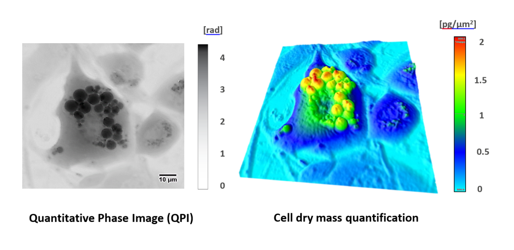
With QPI, not only can cells be visualized with no phototoxicity, but from their intrinsic optic properties the cell dry mass can be directly calculated. The detected phase of light is translated to picograms of cell mass. This allows a very simple and sensitive way for monitoring cell morphology and cell dynamics. Subtle changes in cell populations, rare cell phenotypes, sensitive reactions to drug treatment and many more effects, which remain unseen in the imaging data, can now be explored.
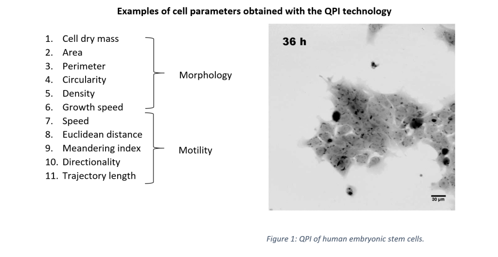
Furthermore, these quantitative images are of high contrast and evince uniform quality which are the crucial requirements for reliable image analysis. Recently, it has been proven that QPI data are highly suitable for object segmentation, cell tracking and advance image analysis using machine learning [2].
New discoveries in cell death research
One of the hallmarks of cancer cells is their ability to avoid programmed cell death. Understanding this phenomenon is one of the main focuses of cancer research and correct cell death type classification is crucial. Predominant types of cell death can be detected by flow cytometry. Nevertheless, the absence of cellular morphology analysis may lead to the misclassification of cell death type. QPI can overcome this phenomenon without the use of fluorescent labels. Researchers from Masaryk University (Brno, Czech Republic) have been using QPI intensively for studies on cell death. Recently, they managed to predict cell death timing using a long-short term memory neuronal network and classify the cell death type as apoptotic or lytic. Importantly, the model was trained solely on QPI data and achieved 75% accuracy [3].
New discoveries in protein condensates
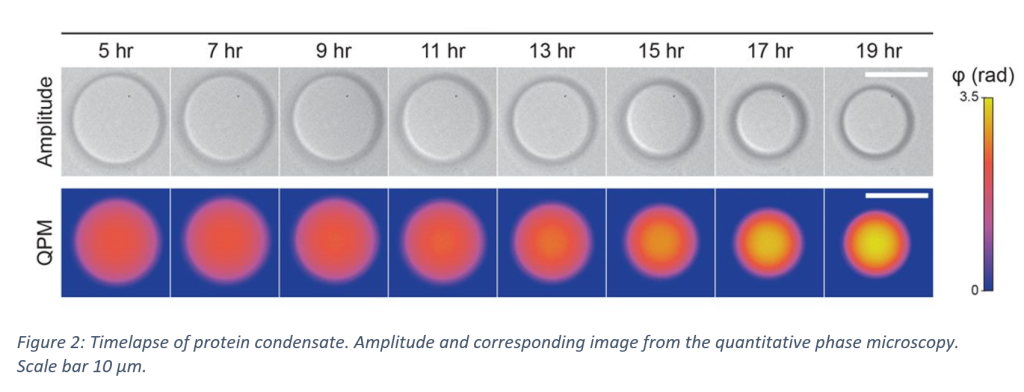
Retrieving the information about the transmitted light wave phase can be used not only for the quantification of cell dry mass, but the obtained phase values can be used to study the composition of biomolecular condensates. These membrane-less and protein-rich cell compartments have been shown to play an important role in biochemical cellular processes. However, their composition, that crucially affects the phase separation mechanism, remains less understood. The research team from MPI (Dresden, Germany) found out that using QPI, the protein concentration and condensate temperature-dependent shape can be determined more precisely and more efficiently when compared with traditionally used methods such as fluorescence microscopy or optical diffraction tomography [4].
QPI brings new possibilities in live-cell imaging and condensate studies, but many more discoveries are yet to come.
Want to know what QPI can do for your research?
Companies like Telight, specialists in quantitative phase imaging, offer professional consultation on QPI and its applicability to specific research. If you have ever struggled with phototoxicity, disturbances during staining or cell segmentation, struggle no more. Assisted sample analysis can be conducted in several places around Europe through the EuroBioimaging platform or via www.telight.eu.

Upcoming webinar
QPI technology and its applications will also be the topic of the upcoming FocalPlane features… webinar. Do not miss your chance to learn more about QPI from the top scientists in the field. Register to join us on 30 June 2022 at 14:30 BST.
References:
[1] Park, Y., Depeursinge, C., & Popescu, G. (2018). Quantitative phase imaging in biomedicine. Nature Photonics 2018 12:10, 12(10), 578–589. https://doi.org/10.1038/s41566-018-0253-x[2] Jo, Y. J., Cho, H., Lee, S. Y., Choi, G., Kim, G., Min, H. S., & Park, Y. K. (2018). Quantitative Phase Imaging and Artificial Intelligence: A Review. IEEE Journal of Selected Topics in Quantum Electronics, 25(1). https://doi.org/10.1109/JSTQE.2018.2859234
[2] Jo, Y. J., Cho, H., Lee, S. Y., Choi, G., Kim, G., Min, H. S., & Park, Y. K. (2018). Quantitative Phase Imaging and Artificial Intelligence: A Review. IEEE Journal of Selected Topics in Quantum Electronics, 25(1). https://doi.org/10.1109/JSTQE.2018.2859234
[3] Vicar, T., Raudenska, M., Gumulec, J., & Balvan, J. (2020). The Quantitative-Phase Dynamics of Apoptosis and Lytic Cell Death. Scientific Reports, 10(1), 1–12. https://doi.org/10.1038/s41598-020-58474-w
[4] McCall, P. M., Kim, K., Fritsch, A. W., Iglesias-Artola, J. M., Jawerth, L. M., Wang, J., Ruer, M., Peychl, J., Poznyakovskiy, A., Guck, J., Alberti, S., Hyman, A. A., & Brugués, J. (2020). Quantitative phase microscopy enables precise and efficient determination of biomolecular condensate composition. BioRxiv, .(.), 2020.10.25.352823. https://doi.org/10.1101/2020.10.25.352823
 (1 votes, average: 1.00 out of 1)