Advertisement

Displaying posts in the category: Default

From Microscope to Macro-Scope: Same Sobek, New Glyphs 

Posted by , on 5 September 2024

Thousands of years ago, the Ancient Egyptians depicted their numerous gods on hieroglyphics. One of which is an amalgamation of crocodile and man – Sobek, the god of the Nile River. These sacred carvings (from the classical Greek “heiros”, sacred; “glyphikos”, carving) have to be deciphered carefully by historians who are far removed from the

BioImage.IO Chatbot: Ready to use and discover!

Posted by , on 21 August 2024

We are delighted to announce the publication of the BioImage.IO Chatbot in Nature Methods this August and its immediate availability for the bioimaging community. This milestone signifies academic recognition and marks the chatbot’s readiness for everyday use in streamlining complex bioimage analysis tasks. Broadening Access to Advanced Bioimage Analysis The BioImage.IO Chatbot, equipped with state-of-the-art

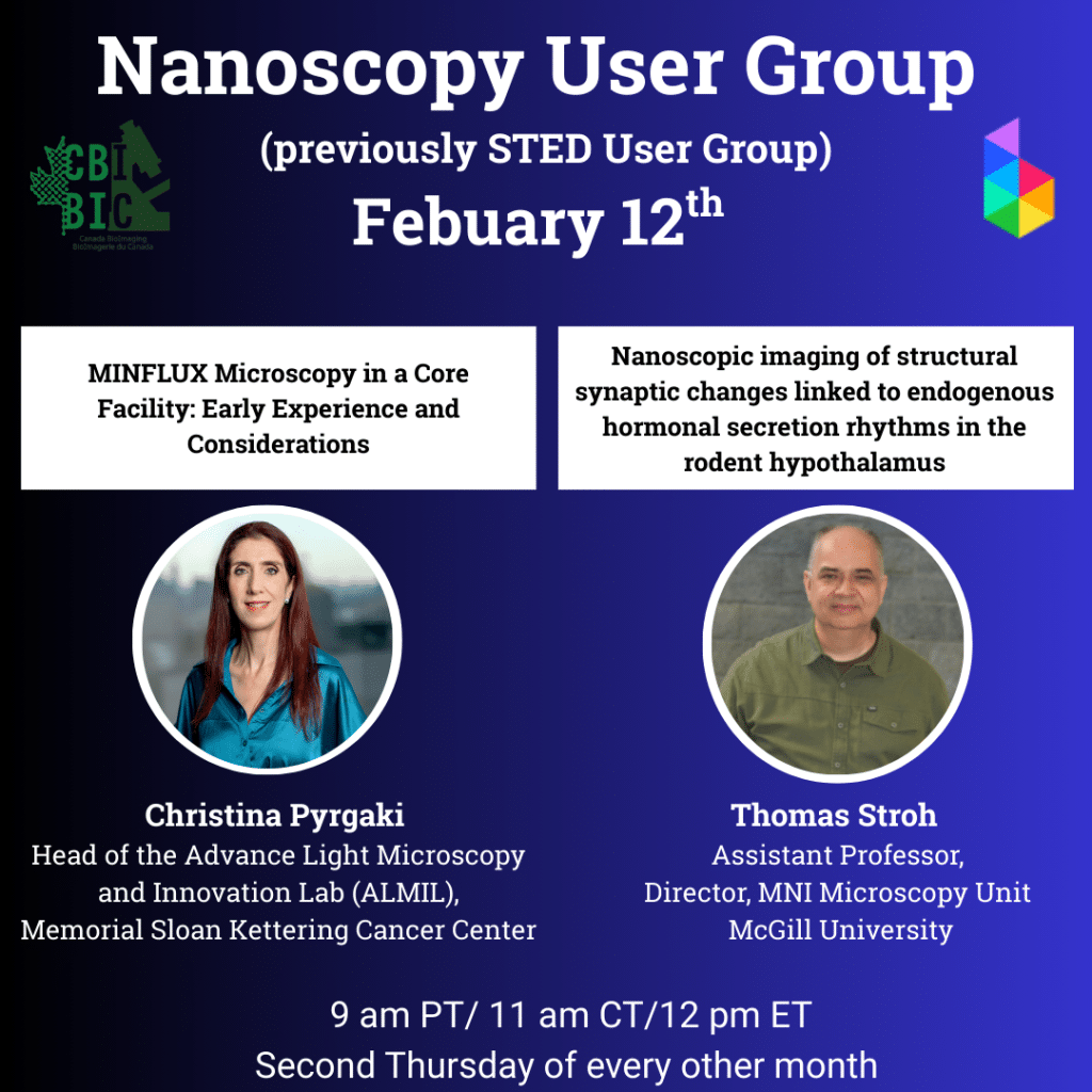
STED User Group (BINA/Canada BioImaging)

Posted by , on 25 July 2024

The next installment of the BioImaging North America and Canada BioImaging STED user group will take place via Zoom on August 8 at 12 pm EST. This user group is targeted at users of all experience levels, and aims to give all members of the community a chance to share technical know-how by facilitating open discussion. Interested participants

Prompt Engineering in Bio-image Analysis

Posted by , on 18 July 2024

Prompt engineering and its importance Communication is key. This saying is not only true in day-to-day life, but also when interacting with Generative Artificial Intelligence, a system able to generate text, images or other output types in response to prompts. In prompt engineering, we use natural language to describe the task that a Large Language

JCS-FocalPlane Training Grants

Posted by , on 15 July 2024

We are delighted to have awarded our first JCS-FocalPlane Training Grants to four early-career researchers who will be attending microscopy training courses over the coming months. Look out for their posts on FocalPlane to discover more about the researchers and the courses that they are attending! The next round of applications for training grants is

AI4Life Event Highlights: A Successful Fusion of Workshops, Hackathon, and Uploathon

Posted by , on 25 June 2024

The recent AI4Life event, hosted at UC3M premises in Madrid, marked a significant milestone in the field of AI and microscopy, drawing a dynamic crowd of approximately 60 participants over five action-packed days. The event was organized by the AI4Life consortium teams of UC3M, KTH, and Euro-BioImaging, It integrated workshops, a hackathon, and an uploathon,

An interview with Juan Quintana

Posted by , on 25 June 2024

MiniBio: Juan Quintana is a Wellcome Trust Fellow and group leader at the Division of Immunology, Immunity to infection, and Respiratory Medicine, the Lydia Becker Institute of Immunology and Inflammation, and the Geoffrey Jefferson Centre for Brain Research at the University of Manchester, UK. The Quintana lab works intersectionally to understand the immunological basis of

Microscopy Training Needs Survey

Posted by , on 20 June 2024

The Royal Microscopical Society (RMS) are currently running a Training Needs Survey, which will be live until 30th July, 2024. Fill in the survey here: https://www.surveymonkey.com/r/66TLZQW In a recent review exercise the RMS established that in 2014 over 30 training courses for different microscopy modalities and techniques were running in the UK. By 2024 this

STED User Group (New Meeting, BINA/Canada BioImaging)

Posted by , on 3 June 2024

BioImaging North America and Canada BioImaging are organizing a new STED user group. The first meeting will take place via Zoom on June 13 at 12 pm EST. This user group is targeted at users of all experience levels, and aims to give all members of the community a chance to share technical know-how by facilitating open discussion.

Education Strategies of Imaging Cores

Posted by , on 1 April 2024

Over the past two decades, high profile research institutions have come to rely on distinct core facilities for much of their data and image analysis. The benefits of the newest technologies are often softened by the costs of purchasing and maintaining such equipment for regular use. For microscopy and imaging, it only made sense that