Welcome to FocalPlane
FocalPlane is a community site for anyone who uses microscopy in their research. It is a place where you can interact with a global community of imaging scientists, engineers, chemists, and bioimage analysts.
FocalPlane is your site and once registered, you are free to share a blog post, add an image to our gallery, post a job advert or put up an event listing.
Create an account to share your story or to stay up-to-date with the latest news.

Recent posts
Microscopy preprints: bioimage analysis
Posted by FocalPlane, on 24 July 2026
JCS snapshot: A ULK1–MTFR1L feedback loop links mitochondrial fission, mitophagy and apoptosis
Posted by FocalPlane, on 22 July 2026
Pearling and directed trafficking as determinants of mitochondrial genome organization and network architecture
Posted by FocalPlane, on 21 July 2026
Featured image with Manomay Pawar
Posted by FocalPlane, on 20 July 2026
Microscopy preprints: applications in biology
Posted by FocalPlane, on 15 July 2026
Compartmentalized Cytoplasmic Tradewinds
Posted by GalbraithLab, on 9 July 2026
Imaging spotlight: VLab4Mic
Posted by FocalPlane, on 7 July 2026

Filter by technique
Filter by research topic
Questions about FocalPlane?
Become a sponsor
Are you interested in becoming a sponsor for FocalPlane?
Our sister sites

The community site for developmental
and stem cell biologists
Image credits
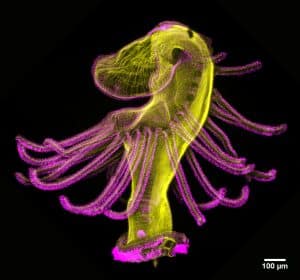
Neuromuscular junctions – Rebecca Simkins
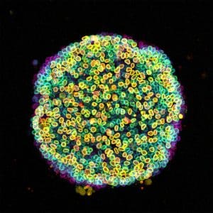
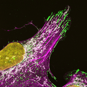
Mitochondria and microtubules – Till Stephan
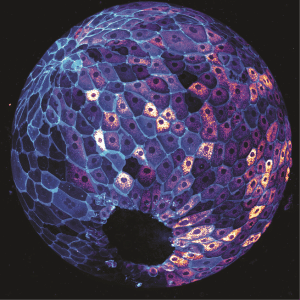
Mammary gland organoid – Oona Paavolainen
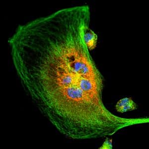
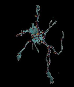
Dopaminergic neuron – Nick Gatford
CLEM HeLa cell – Marie-Charlotte Domart, Chris Peddie
Posts by categories


Microscopy-related articles from our journals
- Toggle-Untoggle – a cell segmentation tool with an interactive user verification interface J Cell Sci 2025 138: jcs264154
- The ratio of Wnt signaling activity to Sox2 transcription factor levels predicts neuromesodermal fate potential Development 2025 152: dev204661
- The short isoform of Tango1 is dispensable for zebrafish survival but is required for skeletal patterning and integrity Biology Open 2025 14: bio062117
Microscopy-related preprint highlights
- When geometry isn’t enough, actin helps plant cells decide where to divide, ensuring robust tissue patterning
- Planar cell polarity protein, Vangl2, goes out of its way to shape the heart through a planar polarity-independent mechanism.
- HAK-actin, U-ExM-compatible probe to image the actin cytoskeleton


Gallery categories
- Bioimage analysisApply
- WidefieldApply
- Confocal microscopyApply
- Spinning disc micros..copyApply
- Super-resolution mic..roscopyApply
- Single plane illumin..ation microscopy (SPIM / Light sheet)Apply
- Total internal refle..ction fluorescence (TIRF) microscopyApply
- Electron microscopyApply
- Multi-photon microsc..opyApply
- Label-free microscop..yApply
- DyesApply
- Genetic probesApply
- Optical manipulationApply
- Functional imagingApply