Featured image with Hoang Anh Le
Posted by FocalPlane, on 5 July 2024
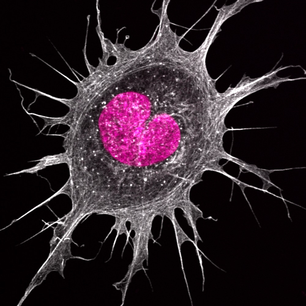
Our featured image, acquired by Hoang Anh Le, shows an Ewing’s sarcoma cancer cell plated on a fibronectin-coated surface. The cell was fixed with 4% formaldehyde and stained using phalloidin (white) and DAPI (in pink). The sample was imaged using a confocal Zeiss 880 microscope with Airyscan.
Find out more about Anh’s research below:
Research career so far: I finished my PhD with Laura Machesky who was then at the Cancer Research UK Scotland Institute (formerly known as the Beatson Institute) working on cancer cell migration and macropinocytosis. I am now a postdoc scientist at University College London. I’m still working on cell migration but using a completely different model system now, the frog embryo. My current research is funded by a Henry Wellcome fellowship looking at how macrophages interact with their surrounding tissue. I still get to use lots of different microscopes though 😉
Current research: My current research theme falls into the realm of a new emerging field called mechanobiology. I use the frog embryo as a model system, looking at how their immune system develops and interacts with their surrounding tissue. The frog embryos are great for mechanical manipulation and measurement, which can be very difficult to do on other model systems. I also still do a lot of imaging, both in vitro and in vivo, though the frog embryos are a bit more challenging to image, but I like a challenge.
Favourite imaging technique/microscope: I love confocal timelapse microscopy. It’s very versatile, and I get to be quite creative with it trying to image the frog embryos. I learn a lot through these trial-and-error processes. I do miss working with cell lines sometimes because it was very easy to image them and they were very easy to maintain as well. But I guess if I don’t go outside of my comfort zone, I won’t learn as much.
What are you most excited about in microscopy currently/in the future? I think it’s definitely the use of AI in image analysis. I’m excited to one day to just be able to type into a console “segment this image and measure the size of the cells” or something like that and the computer just does it for you. I guess people working on natural language processing and AI can help me with that! I’m more of a practical wet lab biologist so if I can just jump straight to the interesting biology question from my images, then I’ll be happy!
 (No Ratings Yet)