Imaging of living cells and tissue is now common in many fields of the life and physical sciences, and is instrumental in revealing a great deal about cellular dynamics and function. It is crucial when performing such experiments that cell viability is at the forefront of any measurement to ensure that the physiological and biological processes that are under investigation are not altered in any way. Many cells and tissues are not normally exposed to light during their life cycle, so it is important for microscopy applications to minimize light exposure, which can cause phototoxicity. To ensure minimal light exposure, it is crucial that microscope systems are optimized to collect as much light as possible.
This can be achieved using superior-quality optical components and state-of-the-art detectors. This Commentary discusses how to set up a suitable environment on the microscope stage to maintain living cells. There is also a focus on general and imaging-platform-specific ways to optimize the efficiency of light throughput and detection.
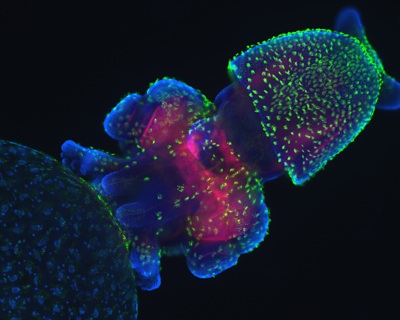
Details about this image 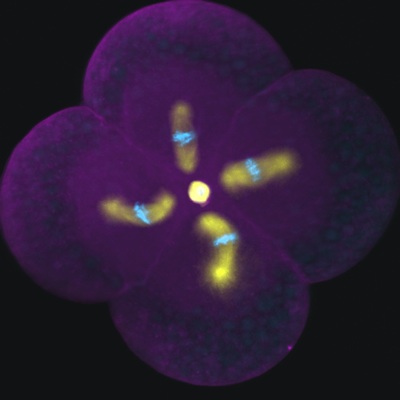
Details about this image 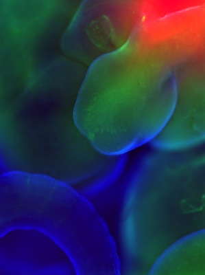
Details about this image 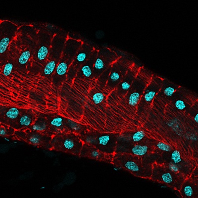
Details about this image
With an efficient optical microscope and a good detector, the light exposure can be minimized during live-cell imaging, thus minimizing phototoxicity and maintaining cell viability. Brief suggestions for useful microscope accessories as well as available fluorescence tools are also presented. Finally, a flow chart is provided to assist readers in choosing the appropriate imaging platform for their experimental systems.