Advertisement

Featured image with Saurabh Chand Sagar

Posted by , on 25 April 2025

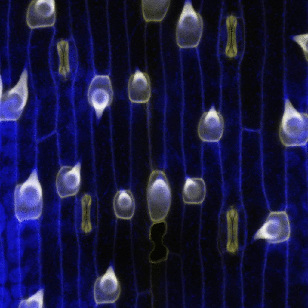
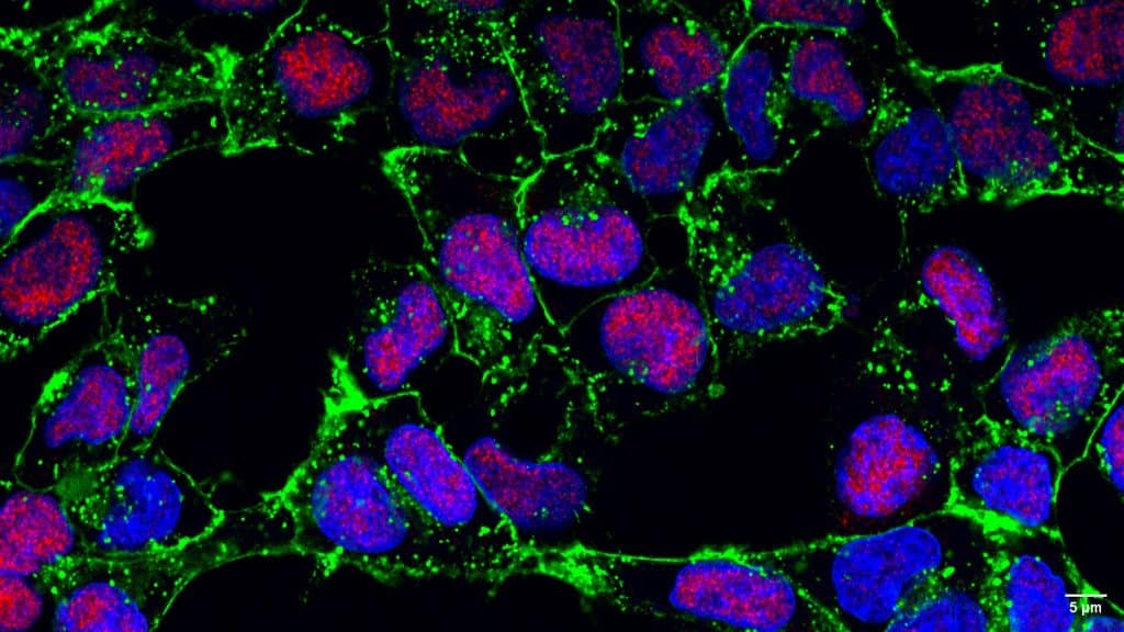
Our featured image, acquired by Saurabh Chand Sagar, presents a laser scanning confocal projection of the Malpighian tubules (MTs) from third instar wandering-stage (118 ± 2 hours post-eclosion) wild-type Drosophila melanogaster larvae. In the image, red denotes F-actin protein, green highlights Discs Large, a protein involved in maintenance of apicobasal polarity, and blue marks the

Microscopy preprints - bioimage analysis

Posted by , on 18 April 2025

Here is a curated selection of preprints published recently. In this post, we focus specifically on bioimage analysis and data management.

FLIMposium - Wednesday 7th May - King's College London

Posted by , on 15 April 2025

The FLIMposium is a one-day symposium dedicated to the latest advances in Fluorescence Lifetime Imaging Microscopy (FLIM). The event will take place on Wednesday, 7th of May, at the Science Gallery, Guy’s Campus, King’s College London. The symposium will feature keynote speakers and invited talks across three main themes: There will also be lightning talks and interactive breakout discussions

Microscopy Meets Medicine: Spirulina-Derived Nanoparticles Take Aim at Cancer

Posted by , on 14 April 2025

This article from the International Journal of Nanomedicine presents a promising new biomimetic strategy that makes use of natural resources as an alternative in cancer therapy. The Researchers have created Spirulina-derived Nanoparticles (sNPs) using a solvent-free process and with the aid of centrifugation and sonication technique. The lysosomes sus-pended averaged an approximate size of 126nm,

Essay competition: Innovative ideas for the future of sustainable events

Posted by , on 11 April 2025

The Company of Biologists is organising an essay competition entitled “Innovative Ideas for the Future of Sustainable Events”. Participants are invited to write an essay of maximum 1000 words to details how your idea will change the way we organise scientific events and will open the door to a new concept of organising events more

Featured image with Subhajit Karmakar

Posted by , on 11 April 2025

Our featured image, acquired by Subhajit Karmakar, depicts a dividing MDA-MB-231 cancer cell that has been stained to visualise the localisation of two key proteins: CK2, a highly oncogenic kinase, and an E3 ligase, a tumour suppressor.

Boston Bioimage Analysis Course (BoBiAC)

Posted by , on 10 April 2025

The Image Analysis Collaboratory and the Core for Imaging Technology & Education at Harvard Medical School are organizing the Boston Bioimage Analysis Course (BoBiAC), a 6-day intensive program offering a hands-on introduction to image processing and analysis on fluorescence microscopy images with Python! If you answered yes to these questions, then this course is for

Image Analysis course with Fiji/ImageJ

Posted by , on 9 April 2025

Online course introducing you to the basics of image analysis, including automatic segmentation, colocalisation, denoising, 3D, etc.

Microscopy preprints - applications in biology

Posted by , on 4 April 2025

Here is a curated selection of preprints published recently. In this post, we share preprints that use microscopy tools to answer questions in biology.

STED Microscopy User Group, April 10, 12pm EDT

Posted by , on 29 March 2025

The next BioImaging North America and Canada BioImaging STED user group will take place via Zoom on April 10 at 12 pm EST. Mathias Delhaye and Jeffrey LeDue will jointly deliver the presentation, “Expanding Insights: Magnified Analysis of the Proteome Across Imaging Platforms”, followed by Q&A. We hope you will join us! This user group is targeted at