Featured image with Jen Silverman
Posted by FocalPlane, on 24 May 2024
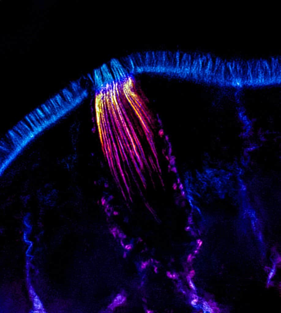
Our featured image, acquired by Jen Silverman, shows a lateral view of a tuft cell taken from a frozen tissue section and imaged using structured illumination microscopy (SIM). F-actin, labelled using phalloidin, is shown in cyan and an actin bundling protein enriched in tuft cells is shown in orange. Reconstruction of the image was performed using Nikon Elements software and post-processing was done using FIJI (ImageJ) and Lightroom (Adobe).
Find out more about Jen’s research below:
Research career so far: I am a currently a 4th year Ph.D. candidate in the Cell and Developmental Biology program at Vanderbilt University in the lab of Dr. Matthew Tyska. Before starting my training, I worked as a research assistant at the Emory Vaccine Center in the lab of Dr. Guido Silvestri, where we worked on antibody targeting in HIV vaccines. This experience jumpstarted my love of cell biology and I began my graduate program looking for ways to incorporate imaging in my research. In the Tyska Lab, we study the structure and formation of the brush border in the small intestinal epithelium using high resolution light and electron microscopy.
Current research: My research is currently focused on tuft cells, a rare, chemosensory cell type in the small intestine. Previous studies found that tuft cells sense parasites and initiate an immune response, aiding in parasite clearance. Despite the quantity of research centered around tuft cell functionality, there is very little known about their basic cell biology. My work has leveraged advanced light and electron microscopy to perform quantitative morphometry of the intestinal tuft cell’s unique cytoskeleton. Insights derived from this work will provide a foundation for understanding of how giant core actin bundles contribute to tuft cell function and the maintenance of intestinal homeostasis.
Favourite imaging technique/ microscope: I enjoy using many microscopes to visualize different aspects of cells, and I appreciate the different strengths and weaknesses of each scope which allows us to capture a wide range of data. While Nikon’s spinning disk confocal is a powerhouse for data collection (and the microscope I logged the most hours on), the transmission electron microscope is my favorite because the clarity and ultrastructural detail of cell structures is mind blowing!
What are you most excited about in microscopy? I am most excited about the development of super-resolution imaging techniques that allow for excellent resolution in three dimensions, even when capturing large samples volumes. Expansion microscopy is one such technique that has a lot of promise for large cytoskeletal structures, such as those found in tuft cells. Separately, I am also very excited about the increasing appreciation for microscopy as a teaching tool and as art. It’s easy to be excited about biology as a scientist, but I love using microscopy to get the public interested too!
 (2 votes, average: 1.00 out of 1)