
Advertisement

Displaying posts with the tag: is_archive

Living Architectures

Posted by , on 30 June 2026

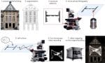
To mark the 40th anniversary of the Musée d’Orsay, the CytoMorpho laboratory was invited to create a performance based on a series of experiments conducted for the occasion. The images produced for this show were real. There were no digital simulations or generative artificial intelligence. They were series of true photographs taken with microscopes.

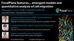
FocalPlane features... cell migration recordings with Juan Manuel Garcia Arcos, Yohalie Kalukula & Daniel J. Cohen

Posted by , on 18 June 2026

In June, we hosted the first webinar in our new series on emergent models and quantitative analysis of cell migration, hosted by Pablo J. Sáez and Valeria Venturini. We heard talks from Juan Manuel García-Arcos, Yohalie Kalukula and Daniel J. Cohen.

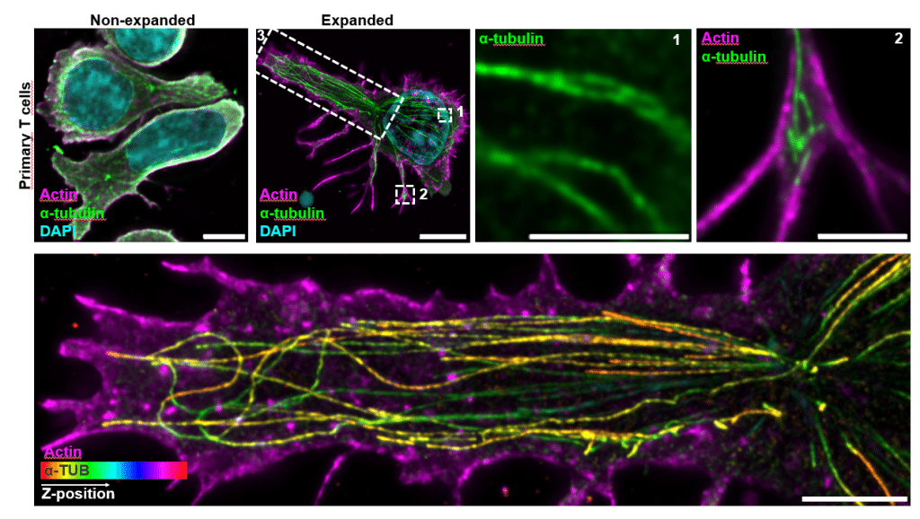
Imaging spotlight: Cryo-ExM reveals the architecture of the human cytotoxic immunological synapse

Posted by , on 15 June 2026

In this behind the paper story, Benita Wolf and colleagues share their research using cryo-expansion microscopy (cryo-ExM) to map the architecture of the human cytotoxic immunological synapse.

Imaging spotlight: the axonal membrane-associated periodic skeleton

Posted by , on 5 March 2026

In this paper highlight, Nick Boyer takes us through the key results and imaging techniques that they used in their research dissecting the axonal membrane-associated periodic skeleton with Rohan Sharma, Christophe Leterrier, Subhojit Roy and colleagues.

Featured image with Jan Martinek

Posted by , on 13 February 2026

Our featured image is a spinning disk fluorescence micrograph, acquired by Jan Martinek, showing an Arabidopsis thaliana root that formed a spiral on a forgotten Petri dish lying flat on the bench. In this orientation, the root cannot grow downward gravitropically, so it keeps turning.

Workshop report: EMBO Course on Image Processing in Cryo-EM

Posted by , on 9 February 2026

Freya Cardozo shares her workshop report from the EMBO course on Imaging Processing in Cryo-EM. Freya's attendance was supported with a JCS-FocalPlane Training Grant.

Imaging spotlight: U-ExM atlas of microbial eukaryotes

Posted by , on 20 January 2026

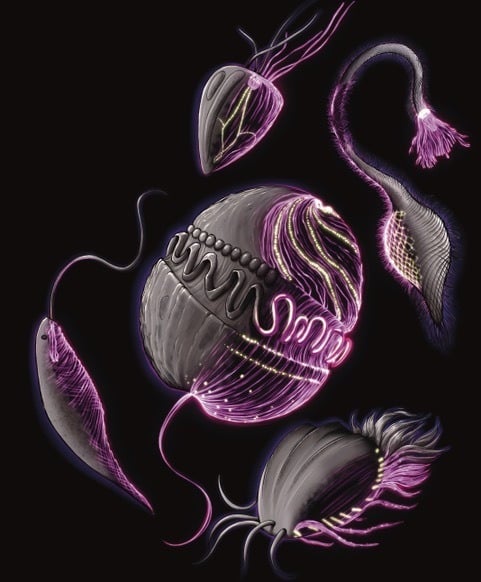
In this paper highlight from Hiral Shah and colleagues, we learn about their ultrastructure expansion microscopy (U-ExM) atlas of over 200 cultured planktonic eukaryotes. This post includes technical advice for ExM users and highlights the open-access resource that they have generated for cytoskeleton or marine microorganism aficionados (and microscopy sciart lovers!)

Microscopy preprints – applications in biology

Posted by , on 19 September 2025

Here is a curated selection of preprints published recently. In this post, we share preprints that use microscopy tools to answer questions in biology.

Featured image with Krystyna Gieniec

Posted by , on 1 August 2025

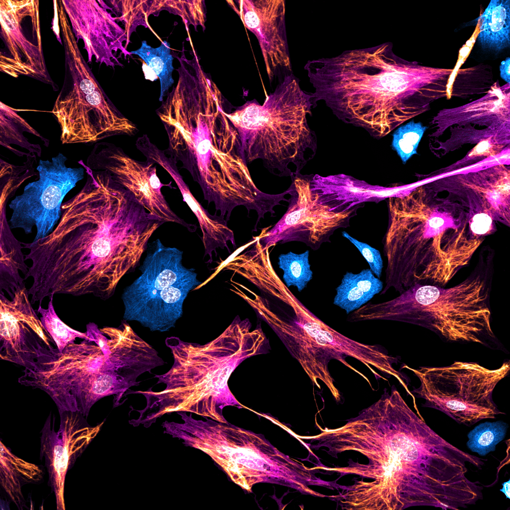
Our featured image, 'Unexpected guests' acquired by Krystyna Gieniec, is a 2D culture of mouse mammary fibroblasts stained for Acta2 (magenta) and Vimentin (gold), with some contaminating epithelial cells stained for pan-Cytokeratin (cyan). The image was acquired using a Leica Stellaris 8 confocal microscope.

Microscopy preprints - applications in biology

Posted by , on 16 May 2025

Here is a curated selection of preprints published recently. In this post, we share preprints that use microscopy tools to answer questions in biology.