Featured image from Lama Khalaily
Posted by FocalPlane, on 27 September 2024
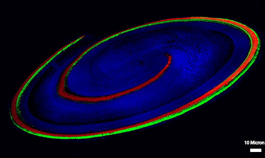
Our featured image, acquired by Lama Khalaily, captures the intricate structure of a mouse cochlea’s hearing organ, highlighting two essential cell types involved in hair cell regeneration. The image reveals four organized rows of sensory hair cells (red), responsible for hearing, intertwined with non-sensory supporting cells (green). These supporting cells play diverse roles in the development, function, survival, and regeneration of sensory epithelia. In the case of hair cell loss (red), the supporting cells (green) can generate new hair cells. However, this ability diminishes after birth. Our research focuses on opening a new possibility for regenerative therapies targeting the conversion of supporting cells into hair cells, offering a potential solution for hearing loss. The image was acquired using a Leica confocal microscope, with post-processing done using ImageJ.
Find about more about Lama’s research below:
Research career so far: I am Lama Khalaily, a Ph.D. student in Prof. Karen Avraham’s lab at Tel Aviv University. I received my B.Sc. in Genetic Engineering and Biotechnology from Jordan University of Science and Technology. I completed my M.Sc. at the University of Haifa in Genetic Regulation and Evolution of Developmental Processes. After finishing my Master’s degree, I worked at the Medical Center – Bruce Rappaport Faculty of Medicine. During this time, I focused on research and clinical projects in human genetics.
Current Research: My Ph.D. studies are focused on understanding the mechanisms of normal trans-differentiation during different developmental and post-natal stages. I am using cochlear explant assays and other high-throughput assays to understand the mechanisms underlying the loss of regenerative potential and the factors that promote trans-differentiation of supporting cells into hair cells. Our research focuses on opening a new possibility for regenerative therapies targeting the conversion of supporting cells into hair cells, offering a potential solution for hearing loss.
Favorite Imaging Technique/Microscope:
Confocal Microscope, Two-Photon Excitation Microscopy, and Super-Resolution Microscopy (SRM)
What Are You Most Excited About in Microscopy? I am most excited about live imaging techniques, where we can see normal development and gain insights into cellular processes in real-time and at molecular resolution. This opens up new possibilities for understanding complex biological systems and disease mechanisms.
 (No Ratings Yet)