Advertisement

Displaying posts with the tag: is_archive

Microscopy preprints: applications in biology

Posted by , on 5 October 2026

Here is a curated selection of preprints published recently. In this post, we share preprints that use microscopy tools to answer questions in biology.

Featured image with Ádám Soós

Posted by , on 25 September 2026

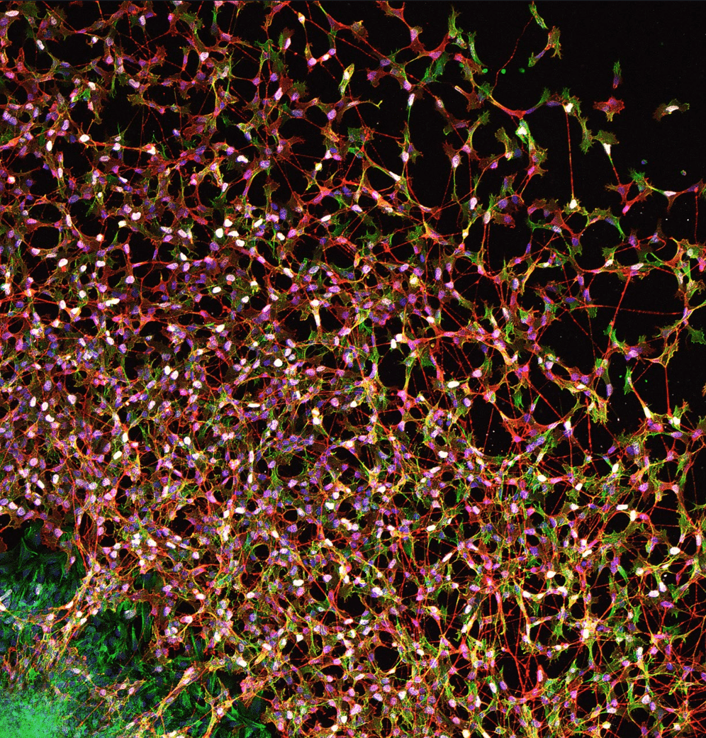
Our featured image, acquired by Ádám Soós is titled “Guided by GDNF: enteric neural crest cells weaving the future gut nervous system.” It shows enteric neural crest-derived cells streaming out from an embryonic day 6 chicken midgut explant, forming an interconnected migratory network on a fibronectin-coated surface. The culture was exposed to 20 ng/ml GDNF,

Featured image with Luz María Fuentealba Pérez

Posted by , on 28 August 2026

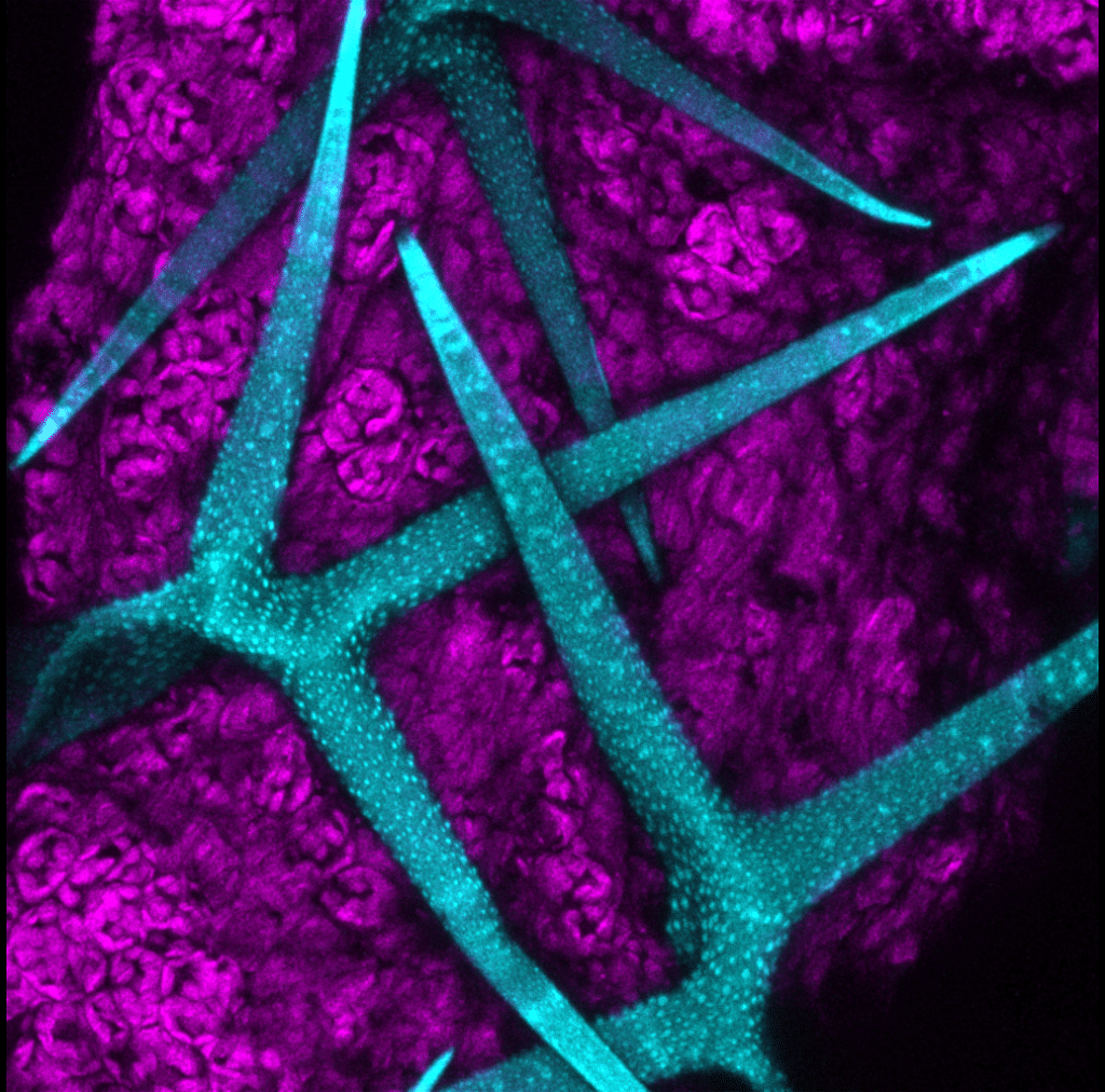
Our featured image, acquired by Luz María Fuentealba Pérez, shows the last breath of Arabidopsis thaliana, the most widely used plant model worldwide.

Microscopy preprints: applications in biology

Posted by , on 29 May 2026

Here is a curated selection of preprints published recently. In this post, we share preprints that use microscopy tools to answer questions in biology.

Featured image with Brittany Carr

Posted by , on 24 April 2026

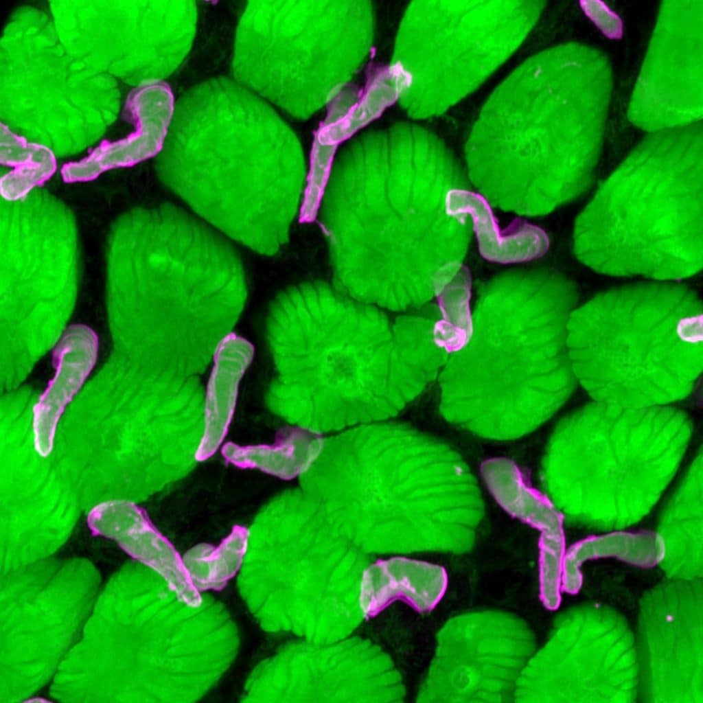
Our featured image, acquired by Brittany Carr, shows the rod and cone photoreceptor outer segments from a tadpole retina. Photoreceptor outer segments are responsible for capturing photons of light and transducing them into chemical signals. Frog rods are large, so this image also shows the incisures, which are invaginations into the outer segment that gives

Featured image with Nat Prunet

Posted by , on 27 March 2026

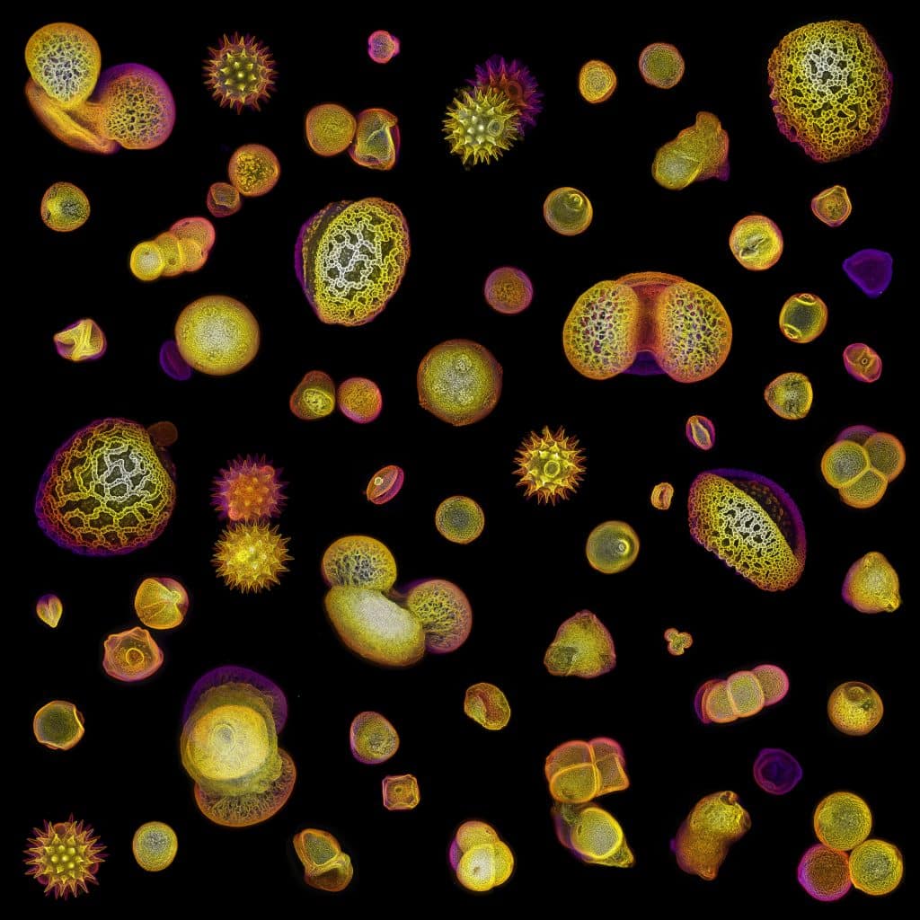
Our featured image from Nat Prunet is a composite image of pollen grains from various plant species. Each pollen grain was imaged separately for red autofluorescence with super-resolution Airyscan on a Zeiss LSM 980 microscope. 3D datasets were processed on Fiji with edge-finding and color-coding for depth.

Workshop report: York Confocal Microscopy course

Posted by , on 3 February 2026

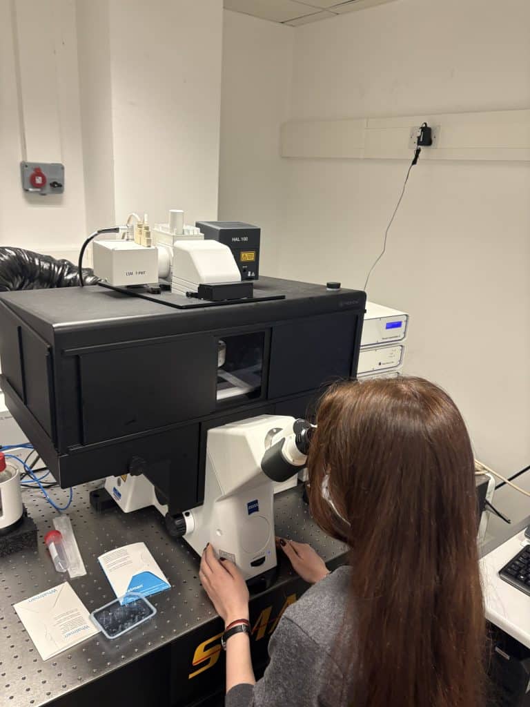
During the four-day Confocal Microscopy 2025 course at the University of York, I gained a better understanding of both the fundamentals of microscopy and more advanced techniques. The daily schedule consisted of the introduction of the relevant topics in the morning, followed by several hands-on practical sessions. In these, we were split into small groups

Featured image with Denise Scuffi

Posted by , on 30 January 2026

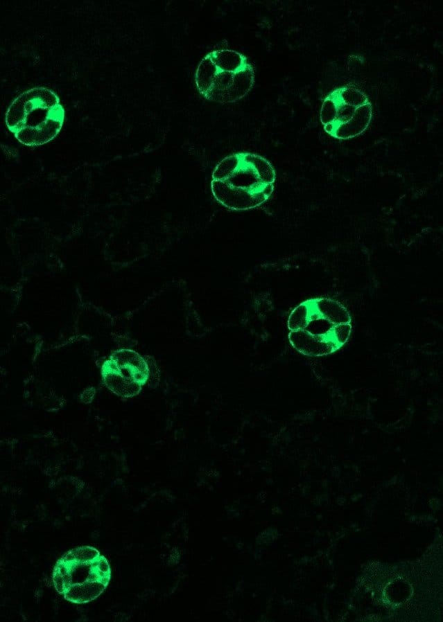
Our featured image, acquired by Denise Scuffi, shows stomata – small pores surrounded by a pair of specialised cells known as guard cells – from Arabidopsis thaliana expressing the fluorescent biosensor roGFP2-Orp1 in the cytosol, which enables in vivo monitoring of intracellular H₂O₂ dynamics.

Microscopy preprints – applications in biology

Posted by , on 23 January 2026

Here is a curated selection of preprints published recently. In this post, we share preprints that use microscopy tools to answer questions in biology.

Imaging spotlight: Initiation of chromosome congression deconstructed by multi-scale live lattice light-sheet and super-resolution microscopy

Posted by , on 16 January 2026

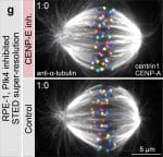
In this highlight, Kruno Vukušić and Iva M. Tolić describe how they use multi-scale live lattice light-sheet and super-resolution microscopy to decipher the earliest steps of chromosome congression.