/* */

Advertisement

Displaying posts in the category: Case studies

FocalPlane features... Samrat Mukhopadhyay with PicoQuant

Posted by , on 31 January 2024

In case you missed ‘FocalPlane features…’, here is the recording of the webinar given by Samrat Mukhopadhyay on 'A deep dive into biomolecular condensates using single-molecule FRET', which we co-hosted with PicoQuant.

Targeted vCLEM of heterogeneous samples

Posted by , on 15 January 2024

by Karel Mocaer* and Paolo Ronchi# *Schwab Team and #EMCF, EMBL Heidelberg, Germany Challenge Studying cells or events within their biological context (i.e. in tissues, ecosystem etc…) is highly relevant. However, volume EM techniques are limited in the volume that can be acquired. Therefore, targeting approaches are needed to characterize specific regions of interest. This

Deciphering cardiac multiscale organization

Posted by , on 11 December 2023

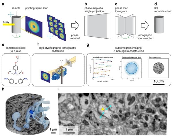
by Sandra Rugonyi and Claudia López Oregon Health & Science University, Portland Oregon, USA Challenge The morphology, microstructure, and ultrastructure of heart tissues is exquisitely organized to optimize cardiac efficiency. In cases of cardiac malformations, known as congenital heart disease (CHD), spatially heterogeneous and abnormal ultrastructural patterns emerge as early as fetal stages that worsen

Upcoming webinar: A Deep Dive into Biomolecular Condensates Using Single-Molecule FRET

Sponsored by PicoQuant, on 22 November 2023

In a brand-new paper, Ashish Joshi and colleagues from the team of Samrat Mukhopadhyay at IISER Mohali take a close look at the molecular events during phase separation of the FUS protein with single-molecule fluorescence methods. If you want to learn more about these techniques and what they revealed about the complex behavior of the

Have you heard of Euro-BioImaging?

Posted by , on 23 March 2023

Euro-BioImaging is a scheme that sounds too good to be true: access to advanced microscopy technologies and the experts that are responsible for them, what’s not to like! The 35 imaging facilities, known as Nodes, that make up the infrastructure of Euro-BioImaging are located across 16 European countries and are accessible to all scientists regardless

How volumeEM (vEM) can help map neuronal circuits

Posted by , on 2 March 2022

by Nadine Randel, University of Cambridge, UK DOI:10.5281/zenodo.6320414 Challenge The nervous system processes sensory information and generates different behavioural outputs accordingly. Synaptic resolution wiring diagrams form a basis for our understanding of how the brain works. Such a connectome provides the synaptic connectivity between all neurons and allows to identify all potential neuronal pathways which

Revealing the spatial regulation of the transcriptome using Split-FISH

Sponsored by Andor, on 9 February 2022

Featured Lab: The Chen Lab, Genome Institute of Singapore  As cells develop and function in tissues, many changes occur not just at the level of RNA transcription, but also at the larger level of spatial organisation of cellular gene expression within the tissue. Studying these changes to RNA profiles at the single-cell level can thus provide

Behind the Screen - behind the scenes of setting up a dynamic screening platform

Posted by , on 22 November 2021

The Cell Biophysics lab has focused on FRET (Forster Resonance Energy Transfer) as it is a powerful technique to investigate dynamic protein-protein interactions, along with studying almost every aspect of cellular signaling with biosensors. FRET is detected either by ratioing the  intensities of the FRET donor and acceptor,  or by FLIM (Fluorescence Lifetime Imaging ;

Asymmetric cell divisions in 3 dimension stem cell colonies

Posted by , on 27 September 2021

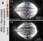
When looking at the development of a multicellular organism, for example a human, the first striking feature is the progressive increase in cell numbers due to successive divisions, from one single tiny cell 80 mm in diameter to a 3.5 kg ball of organised cells forming a new-born baby. The way cells divide, in particular

SRRF-Stream+ Super-Resolution Microscopy Accessible to All

Sponsored by Andor, on 12 August 2020

Fast, reliable & live-cell compatible Super-Resolution Science has limits imposed by the laws of physics that constrain discoveries and the advance of knowledge. In microscopy, up until the beginning of the XXI century, the diffraction limit of light was an unbreakable barrier. This law of physics imposes that two points could not be resolved (clearly