Featured image
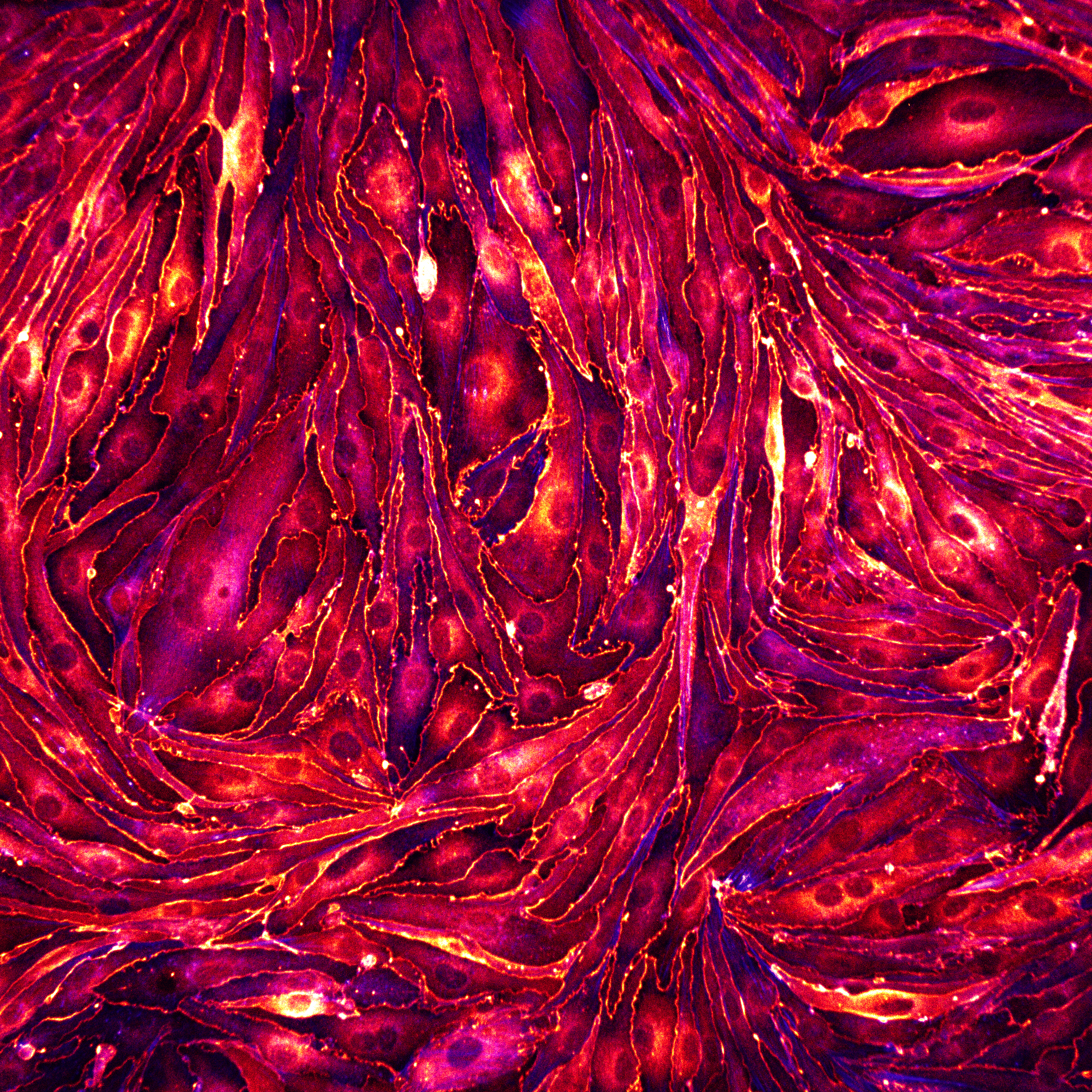
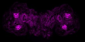
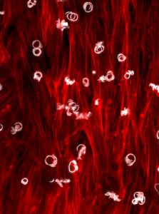
Our featured image, acquired by Rohit Nautiyal, shows a monolayer of brain endothelial cells seeded on a glass substrate coated with fibronectin. The cells were fixed and stained for ZO-1 (LUT- Red Hot) to visualise the tight junctions and actin (LUT-blue). The image was captured with a spinning disk confocal microscope and post-processed with Fiji software.
The copyright for all the images in our gallery remains with the author unless otherwise stated. Please contact the author if you would like to use an image for any purpose.
Gallery
Post an image
Log in or sign up to post an image to the FocalPlane gallery.
Filter by
- WidefieldApply
- Bioimage analysisApply
- Confocal microscopyApply
- Spinning disc micros..copyApply
- Super-resolution mic..roscopyApply
- Single plane illumin..ation microscopy (SPIM / Light sheet)Apply
- Total internal refle..ction fluorescence (TIRF) microscopyApply
- Electron microscopyApply
- Multi-photon microsc..opyApply
- Label-free microscop..yApply
- DyesApply
- Genetic probesApply
- Optical manipulationApply
- Functional imagingApply