Advertisement

Featured image with Omkar Joshi

Posted by , on 14 March 2025

Our featured image, acquired by Omkar Joshi, showcases a U2OS cell immunostained for paxillin (green), TOMM20 (mitochondria, white), and F-actin (labelled using SiR-Actin, magenta), with DAPI (yellow) marking the nucleus.

Featured image with Anthony Dornan and Fara van der Schans

Posted by , on 28 February 2025

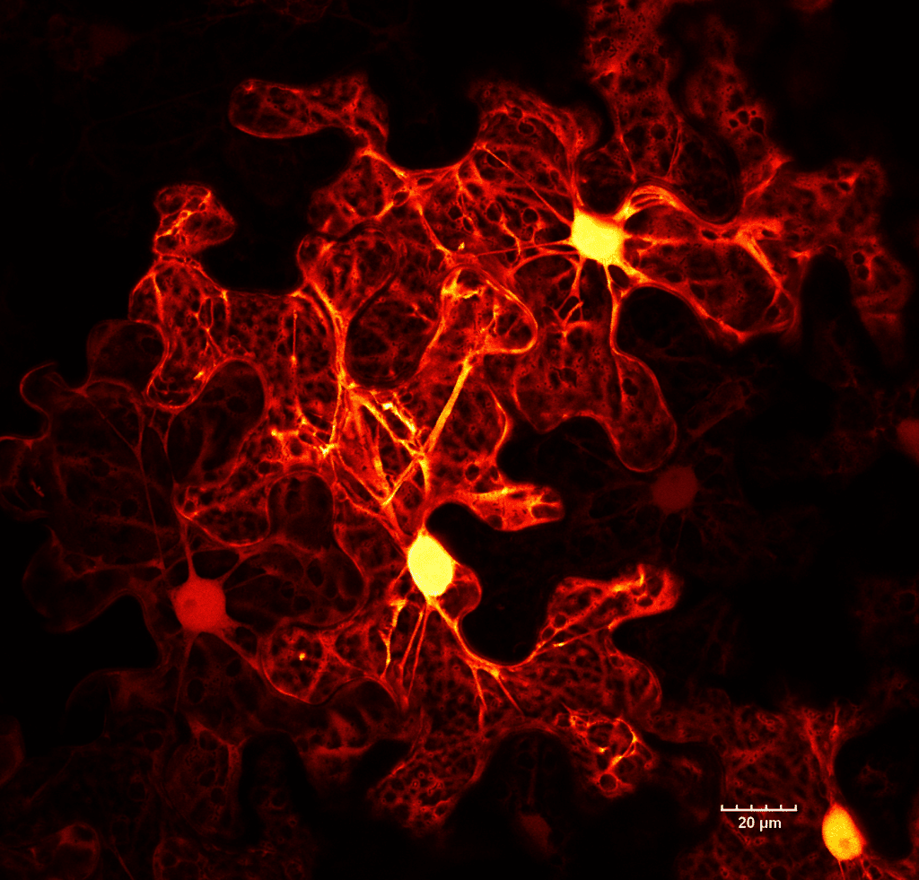
Our featured image, acquired by Anthony Dornan and Fara van der Schans, showcases the perinuclear distribution of mitochondria in the highly respiratory human pancreatic ductal adenocarcinoma (PDAC) tumour cells (Panc-1) stained with the mitochondrial stain TMRE (thermal LUT), the mitochondria are associated with the nuclei, stained with DAPI (cyan). The live cell image is a maximum intensity Z-projection generated

Featured image with Amy Engevik

Posted by , on 14 February 2025

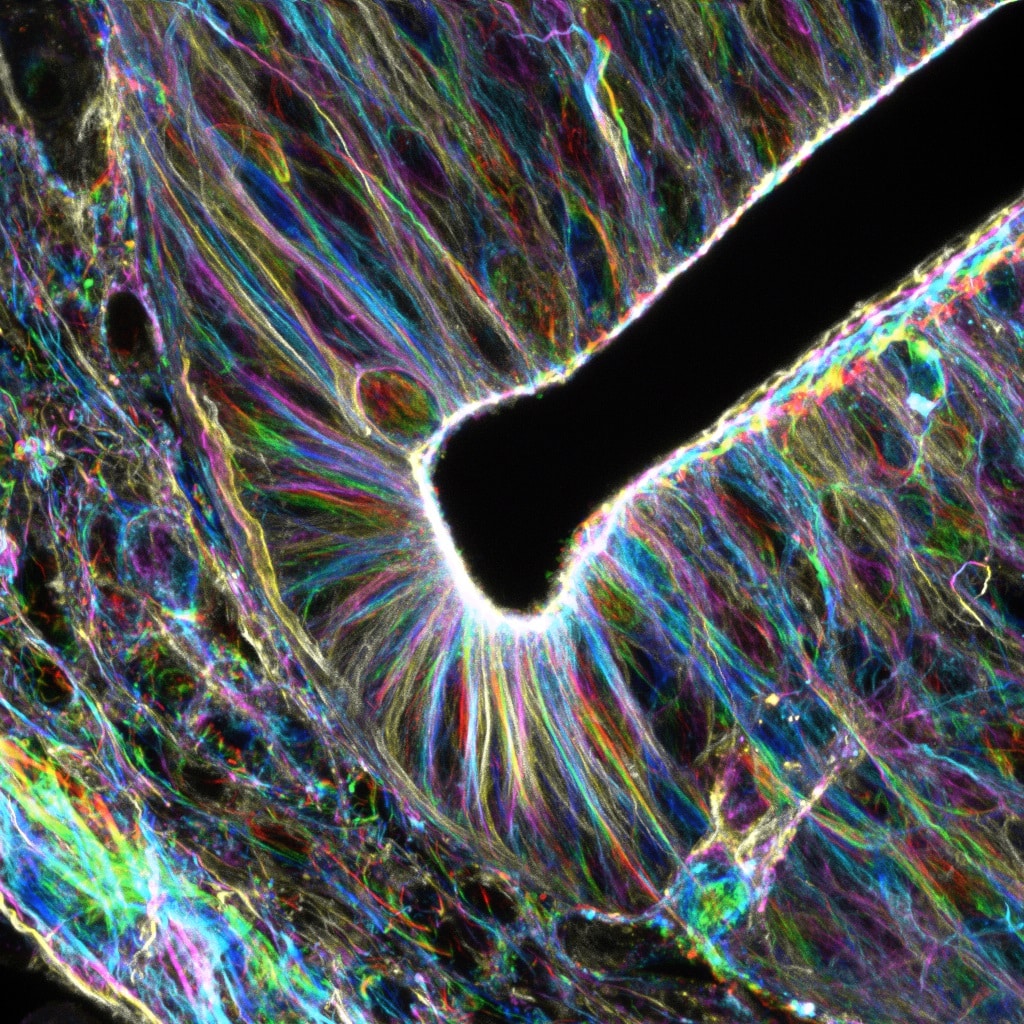
Our featured image, acquired by Amy Engevik, shows a differentiated intestinal organoid from a germline Myosin 5b knockout mouse.

Featured image with Ellen C. O'Shaughnessy

Posted by , on 31 January 2025

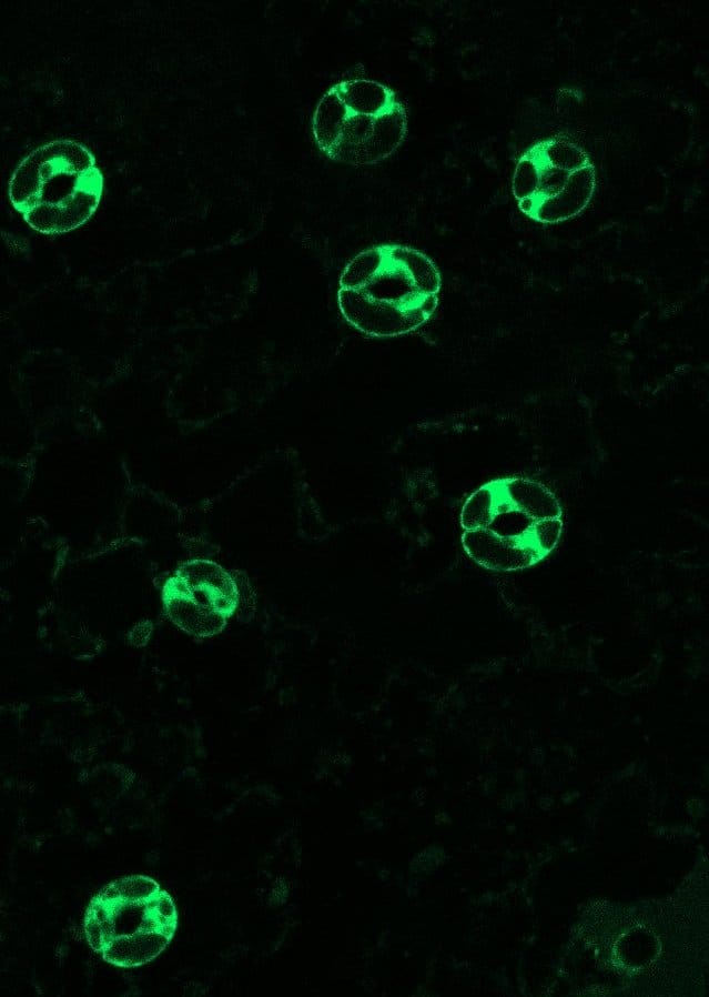
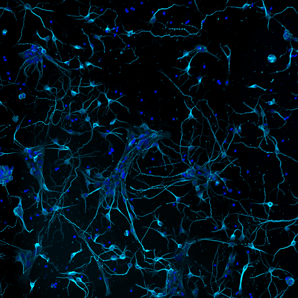
Our featured image shows the locations of exocytic hotspots in a developing cortical neuron.

Featured image with the Hughes lab

Posted by , on 20 December 2024

Our featured image, acquired by Sanja Sviben, is an immuno-scanning electron micrograph of a primary cilium, taken from an intact human pancreatic islet. The cilium is a microtubule-based structure and is seen here labeled with colloidal gold for acetylated alpha tubulin.

Featured image with Jiwon Lee

Posted by , on 6 December 2024

Our featured image, acquired by Jiwon Lee, is a 3D rendering model of Plasmodium falciparum showing the complex organisation of cellular organelles within the malaria parasite. Using a focused ion beam scanning electron microscope (FIB-SEM), a series of high resolution 2D images were acquired. The image stack was aligned and each organelle was segmented in

Featured image with Md Hashim Reza

Posted by , on 22 November 2024

Our featured image, prepared by Md Hashim Reza, is an Andy Warhol-inspired illustration of a Candida albicans cell, post-expansion and labelled with BodipyTR, highlighting features of internal membrane organisation.

Featured image with Jyoti Maddhesiya

Posted by , on 8 November 2024

Our featured image, acquired by Jyoti Maddhesiya, is a laser scanning confocal image of H9c2 cardiomyoblast cells. The green shows BMP7 in the cytoplasm, the blue highlights the nuclei and the cytoskeleton protein F-actin is labelled in red. The cells were transfected with a BMP7 construct and fixed and labelled 48 hours post transfection. The

Featured image with… our JCS cover competition winner Antara Chakraborty

Posted by , on 30 October 2024

We are delighted to announce that our cover competition winner is Antara Chakraborty. Antara’s winning image ‘Breaking symmetry’ is the cover image for Journal of Cell Science’s Special Issue: Imaging Cell Architecture and Dynamics; you can explore the table of contents here. The image shows a mouse embryonic fibroblast labelled with phalloidin (actin – green),

Featured image with David Gaboriau

Posted by , on 11 October 2024

Our featured image, acquired by David Gaboriau, shows human fibroblasts with mitochondria (Tom20) in yellow, alpha tubulin in pink and actin in grey. It’s a 3D-SIM image taken on a Zeiss Elyra PS.1, reconstructed in Zen and presented as a maximum intensity projection. Find out more about David’s research below: Research career so far: I’m originally a