Advertisement

Displaying posts with the tag: is_archive

An interview with Kildare Miranda

Posted by , on 10 May 2022

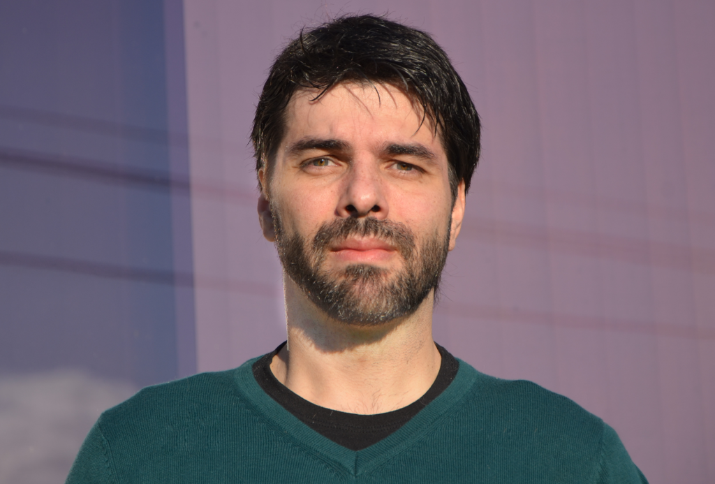
Kildare Miranda is the director of the Advanced Microscopy Unit and vice-director of the National Centre for Structural Biology and Bioimaging (CENABIO), Professor of Biophysics and Cell Biology and Head of the Cell Biology and Parasitology Department of the Biophysics Institute at the Federal University of Rio de Janeiro, and holds several CNPq fellowships. He

An interview with Moara Lemos

Posted by , on 3 May 2022

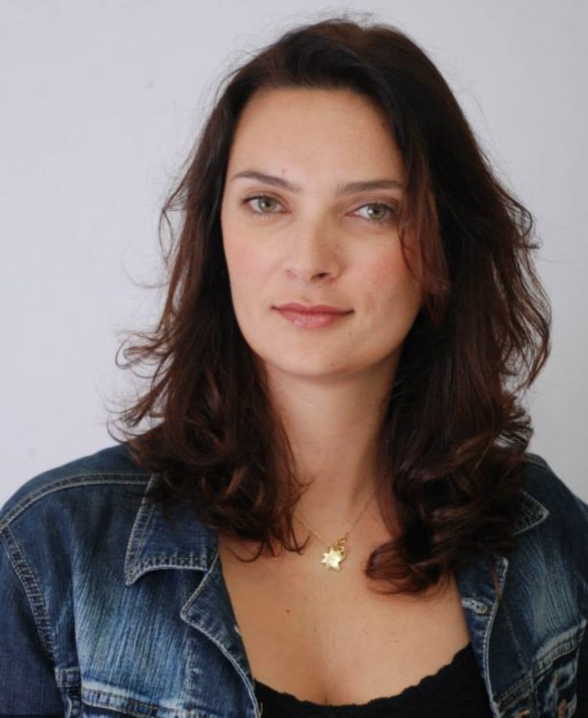
Moara Lemos is currently a permanent researcher at Institut Pasteur in France, where she has helped establish cryo-correlative light and electron microscopy and cellular cryo-tomography for different biological systems. She began her career (BSc and MSc) at the Federal University of Juiz de For a, working on trypanosomes of wild animals. She later did her

An interview with Marcel Cunha

Posted by , on 26 April 2022

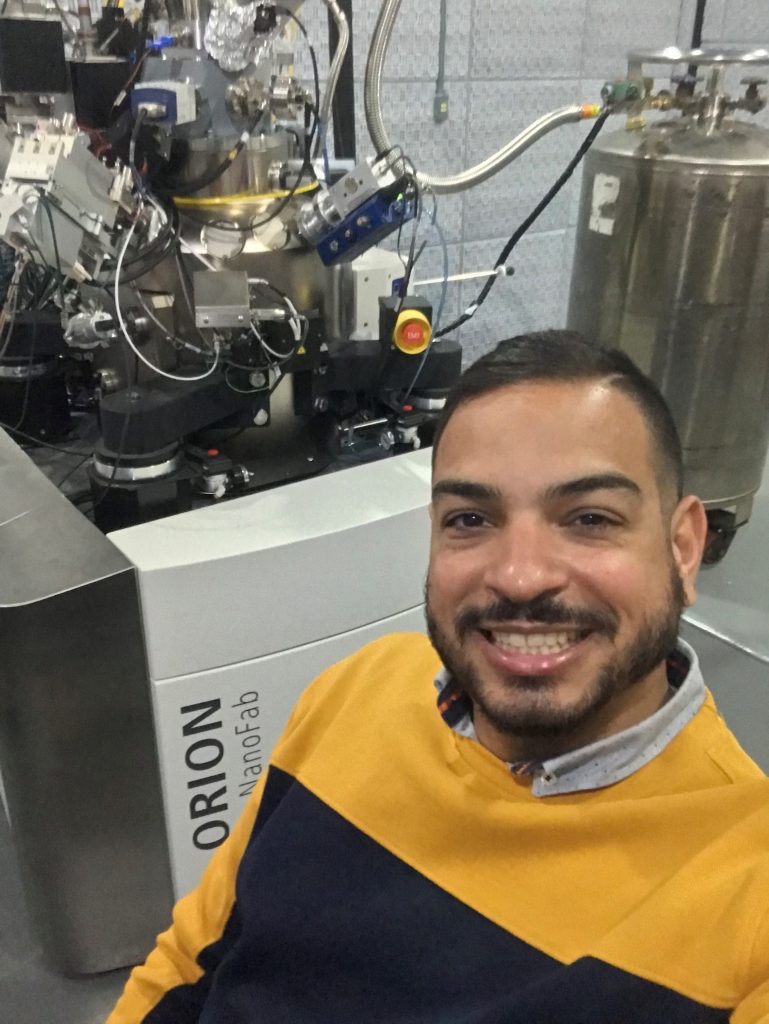
Marcel Cunha is currently an adjunct professor at the Federal University of Rio de Janeiro (UFRJ). He started his career during a BSc degree in Microbiology and Immunology at UFRJ. He then joined Instituto de Biofisica Carlos Chagas Filho (UFRJ) to do his MSc and PhD studies. Later he did his postdoc in Institut Curie

An interview with Victor Midlej

Posted by , on 12 April 2022

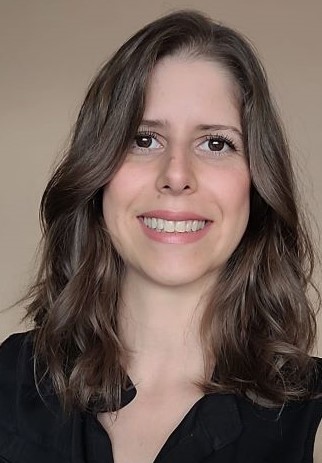
Dr. Victor Midlej is a group leader at Fundação Oswaldo Cruz, working at the interface of microscopy and parasitology, focusing on cell biology, ultrastructure and chemotherapy. He has worked as a microscopist since early in his studies in 2003. Victor began his career (MSc and PhD) at the Universidade Federal do Rio de Janeiro, in

An interview with Lia Medeiros

Posted by , on 5 April 2022

Lia Medeiros is an associate staff scientist at Carlos Chagas Institute, Fiocruz (Oswaldo Cruz Foundation, Brazil), and is head of the Electron Microscopy Facility at the same institution. She obtained her Ph.D. from the Federal University of Rio de Janeiro, Brazil. Her Ph.D. research involved the ultrastructural characterization of the interactions between Plasmodium parasites and

An interview with Leandro Lemgruber

Posted by , on 15 March 2022

Leandro Lemgruber is the facility manager of the Glasgow University Imaging facility in Scotland, UK, and has been in this role since 2015. He has worked in several capacities as a microscopist in Brazil, Italy, Germany, the USA and the UK. Leandro began his work in microscopy and parasitology since his undergraduate degree. He did

How volumeEM (vEM) can help map neuronal circuits

Posted by , on 2 March 2022

by Nadine Randel, University of Cambridge, UK DOI:10.5281/zenodo.6320414 Challenge The nervous system processes sensory information and generates different behavioural outputs accordingly. Synaptic resolution wiring diagrams form a basis for our understanding of how the brain works. Such a connectome provides the synaptic connectivity between all neurons and allows to identify all potential neuronal pathways which

FocalPlane features... CLEM

Posted by , on 31 January 2022

In case you missed ‘FocalPlane features…’ this month, here is the recording of the webinar given by Yannick Schwab (EMBL Heidelberg), Ori Avinoam (Weizmann Institute of Science) and Alexandra Pacureanu (ESRF The European Synchrotron) on Monday 17 January 2022.

Meet the people behind Volume EM community (part 2)

Posted by , on 28 December 2021

In this second post, we continue talking with some of the scientists involved in the Volume EM initiative. If you missed the first part of these series of interview, you can find it here. Raffa Carzaniga and Errin Johnson, Training Working Group What are the aims of your WG? The main aim of the vEM

Meet the people behind Volume EM community (part 1)

Posted by , on 21 December 2021

Volume Electron Microscopy or volume EM (vEM) is a relatively new term that brings together several recently developed imaging approaches that use scanning and transmission electron microscopy (SEM and TEM) to allow the interrogation of cell and tissue ultrastructure in 3D, at μm to mm volume scales and nm resolutions.​ In this blog series ‘volume