Advertisement

Displaying posts with the tag: is_archive

Featured image with Ludovica Altieri

Posted by , on 15 August 2025

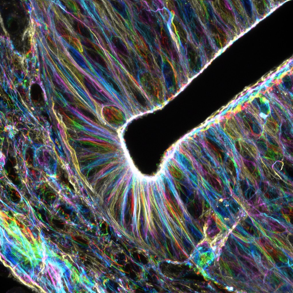
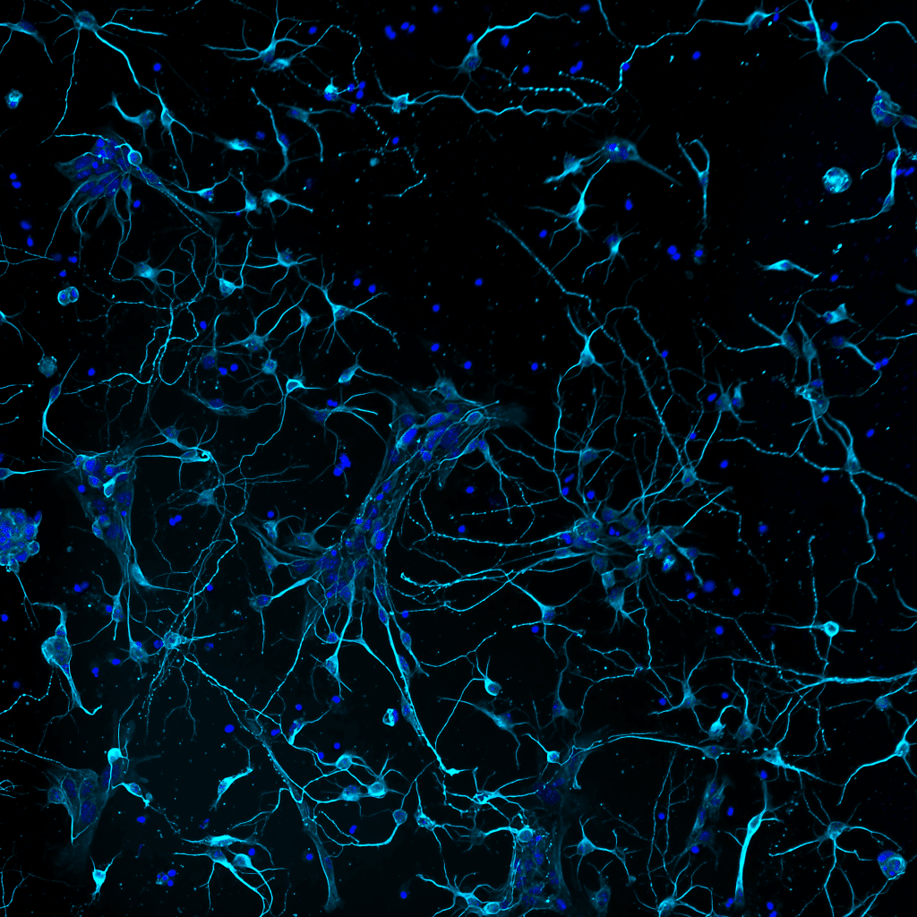
Our featured image, acquired by Ludovica Altieri, shows murine primary cortical neurons developing interconnections. The cells are stained for neuronal tubulin (TUJI antibody, cyan) and DNA (DAPI, blue). Cultures were prepared by dissecting cortices from mouse embryos (E18), then mechanically dissociating single cells for plating. Explanted primary neurons placed in culture regress several stages back,

Featured image with Krystyna Gieniec

Posted by , on 1 August 2025

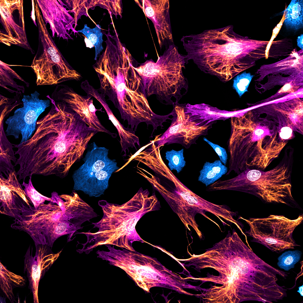
Our featured image, 'Unexpected guests' acquired by Krystyna Gieniec, is a 2D culture of mouse mammary fibroblasts stained for Acta2 (magenta) and Vimentin (gold), with some contaminating epithelial cells stained for pan-Cytokeratin (cyan). The image was acquired using a Leica Stellaris 8 confocal microscope.

Featured image with Maik Bischoff

Posted by , on 18 July 2025

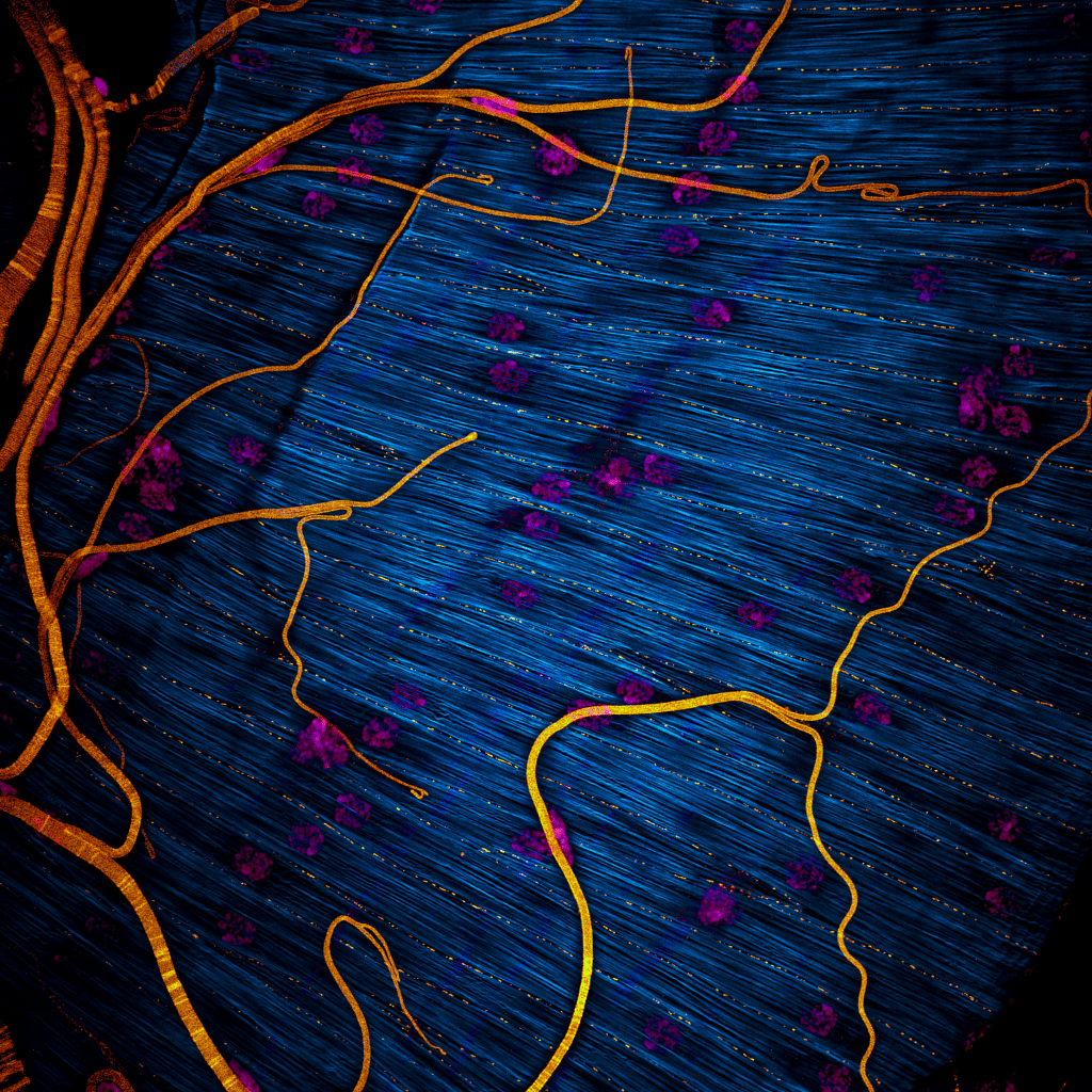
Our featured image, acquired by Maik Bischoff, captures a portion of the testis from the fruit fly Drosophila hydei. Like all insect testes, it is enveloped by a muscle layer. F-actin was stained using phalloidin, making the muscle fibers visible, while DAPI staining revealed the multiple nuclei within each syncytial myotube. An antibody against Drosophila

Featured image with Seth Agyei Domfeh

Posted by , on 20 June 2025

Our featured image, acquired by Seth Agyei Domfeh, shows the nuclear localisation of interferon regulatory factor 9 (IRF9) in human hepatoma (HepG2) cells stimulated with interferon α (IFN-α). HepG2 cells were cultured on a poly-D-lysine-coated coverslip for 24 hours and stimulated with 1000 IU/ml IFN-α. At 24 hours post-stimulation, the cells were fixed using 4%

Featured image with Rosa L. Salgado García

Posted by , on 6 June 2025

Our featured image, acquired by Rosa L. Salgado García, shows a histological section of female (above) and male (below) gonads at an advanced maturation stage of the simultaneous hermaphroditic scallop Nodipecten subnodosus. The image of the gonadic tissue was obtained using a microscope (Leica DM4B) connected to a digital camera (Leica, DMC2900) using LAS V

Featured image with Andrew Octavian Sasmita

Posted by , on 23 May 2025

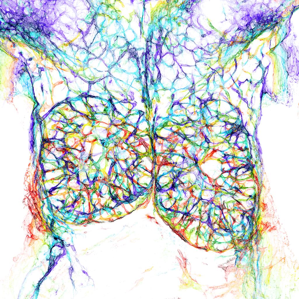
Our featured image, acquired by Andrew Octavian Sasmita, depicts a single brain hemisphere of the 5xFAD mouse model of Alzheimer's disease (AD) at different spatial scales.

Featured image with Rohit Nautiyal

Posted by , on 9 May 2025

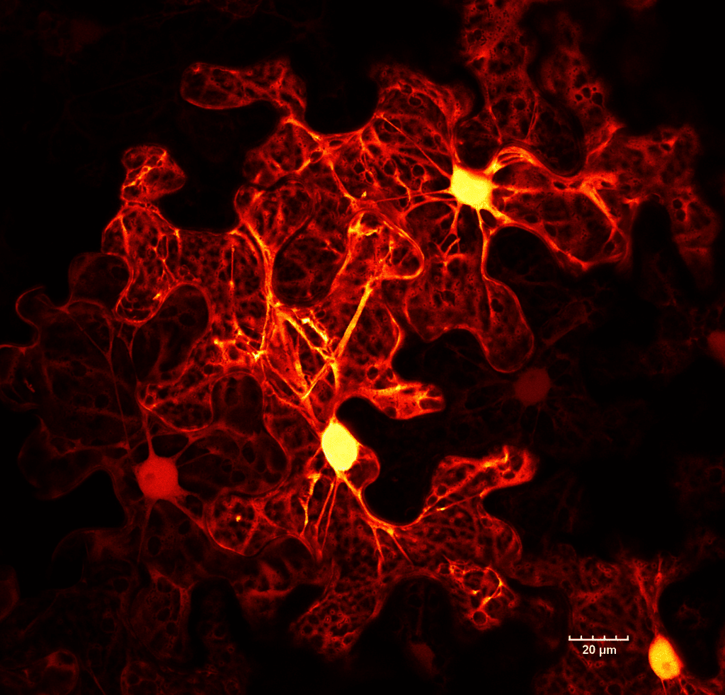
Our featured image, acquired by Rohit Nautiyal, shows a monolayer of brain endothelial cells seeded on a glass substrate coated with fibronectin. The cells were fixed and stained for ZO-1 (LUT- Red Hot) to visualize the tight junctions and actin (LUT-blue). The image was captured with a spinning disk confocal microscope and post-processed with Fiji

Featured image with Saurabh Chand Sagar

Posted by , on 25 April 2025

Our featured image, acquired by Saurabh Chand Sagar, presents a laser scanning confocal projection of the Malpighian tubules (MTs) from third instar wandering-stage (118 ± 2 hours post-eclosion) wild-type Drosophila melanogaster larvae. In the image, red denotes F-actin protein, green highlights Discs Large, a protein involved in maintenance of apicobasal polarity, and blue marks the

Featured image with Subhajit Karmakar

Posted by , on 11 April 2025

Our featured image, acquired by Subhajit Karmakar, depicts a dividing MDA-MB-231 cancer cell that has been stained to visualise the localisation of two key proteins: CK2, a highly oncogenic kinase, and an E3 ligase, a tumour suppressor.

Featured image with Omkar Joshi

Posted by , on 14 March 2025

Our featured image, acquired by Omkar Joshi, showcases a U2OS cell immunostained for paxillin (green), TOMM20 (mitochondria, white), and F-actin (labelled using SiR-Actin, magenta), with DAPI (yellow) marking the nucleus.