Advertisement

Displaying posts with the tag: is_archive

Microscopy preprints - applications in biology

Posted by , on 21 February 2025

Here is a curated selection of preprints published recently. In this post, we share preprints that use microscopy tools to answer questions in biology.

Microscopy preprints - bioimage analysis

Posted by , on 29 November 2024

Here is a curated selection of preprints published recently. In this post, we focus specifically on bioimage analysis and data management.

Microscopy preprints - applications in biology

Posted by , on 15 November 2024

Here is a curated selection of preprints published recently. In this post, we share preprints that use microscopy tools to answer questions in biology.

Featured image with Ciarán Butler Hallissey

Posted by , on 13 September 2024

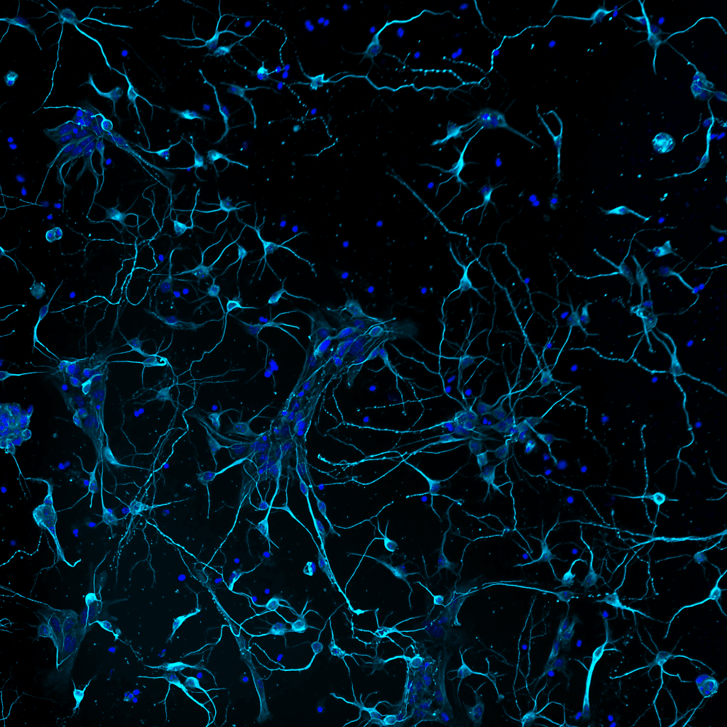
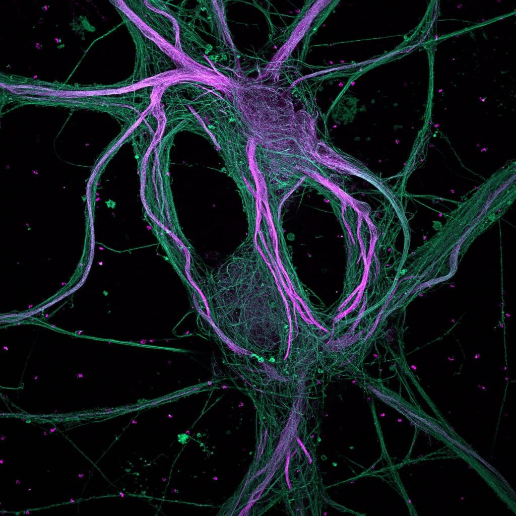
Our featured image, acquired by Ciarán Butler Hallissey, is a maximum-intensity projection of rat hippocampal neurons processed with ultrastructure expansion microscopy (U-ExM) and captured with a spinning disk confocal microscope. Find out more about the image and Ciarán’s research below. More about the image: Expansion microscopy physically increases the size of your sample to improve

Featured image with Shivangi Verma

Posted by , on 30 August 2024

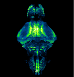
Our featured image, acquired by Shivangi Verma, gives us a peek into the brain of a zebrafish. It depicts neurons in the whole brain of a larva expressing GCaMP6f, a fluorescent reporter of neuronal activity, using a pan-neuronal promoter. A 10-day old transparent homozygous albino larva was embedded in agarose and imaged using ZEISS Lightsheet

Microscopy preprints – applications in biology

Posted by , on 23 August 2024

Here is a curated selection of preprints published recently. In this post, we share preprints that use microscopy tools to answer questions in biology.

Featured image with Upasana Gupta

Posted by , on 2 August 2024

Our featured image, acquired by Upasana Gupta, represents the cellular architecture of zebrafish (larva) eye. The tissue was prepared by paraformaldehyde fixation of a 4-day post fertilisation larva, followed by staining with Hoechst nuclear stain. The sample was imaged and processed using Leica SP8 confocal microscope and Leica software at the Bio-imaging facility at the

Featured image with Theresa Wiesner

Posted by , on 21 June 2024

Our featured image, acquired by Theresa Wiesner, shows a neuron in dissociated hippocampal culture and was imaged using highly inclined thin illumination microscopy (HiLO). The neuron is transfected with a neurotransmitter (glutamate) sensor and shown in orange. The image was acquired using Nikon Elements software / microscope and post-processing was done using FIJI (ImageJ). Find

Featured image with Nick Gatford

Posted by , on 16 November 2023

Our featured image shows a single human dopaminergic neuron generated from a human stem cell, acquired via super-resolution Airyscan confocal microscopy at the University of Oxford Micron facility by Nick Gatford. We caught up with Nick to find out about his research and what he is excited about in microscopy.