Advertisement

Displaying posts with the tag: is_archive

Featured image with Anthony Dornan and Fara van der Schans

Posted by , on 28 February 2025

Our featured image, acquired by Anthony Dornan and Fara van der Schans, showcases the perinuclear distribution of mitochondria in the highly respiratory human pancreatic ductal adenocarcinoma (PDAC) tumour cells (Panc-1) stained with the mitochondrial stain TMRE (thermal LUT), the mitochondria are associated with the nuclei, stained with DAPI (cyan). The live cell image is a maximum intensity Z-projection generated

Featured image with Theresa Wiesner

Posted by , on 21 June 2024

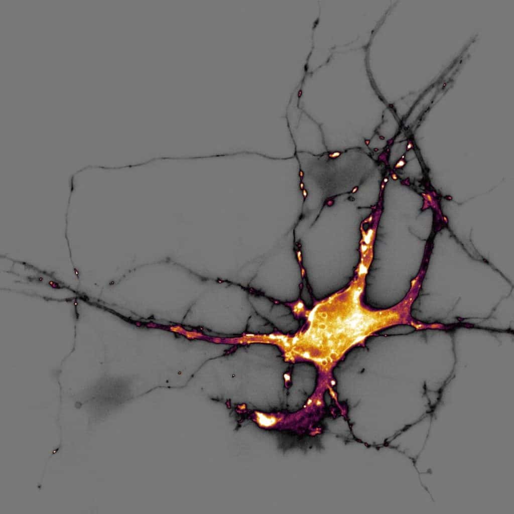
Our featured image, acquired by Theresa Wiesner, shows a neuron in dissociated hippocampal culture and was imaged using highly inclined thin illumination microscopy (HiLO). The neuron is transfected with a neurotransmitter (glutamate) sensor and shown in orange. The image was acquired using Nikon Elements software / microscope and post-processing was done using FIJI (ImageJ). Find

A general strategy to develop fluorogenic polymethine dyes for bioimaging

Posted by , on 6 December 2023

by Annabell Martin and Pablo Rivera-Fuentes. Fluorescence imaging is an invaluable tool to study biological processes and further progress depends on the development of novel advanced fluorescent probes. Excitingly, using polymethines as a scaffold for fluorogenic dyes, we have opened up a lot of new avenues for bioimaging.

Technology Highlights - Expansion Microscopy

Posted by , on 29 August 2023

Interview with Ana Agostinho and Steven Edwards, at the Advanced Light Microscopy facility at SciLifeLab in Sweden. We are today talking about Expansion Microscopy imaging. Please provide a short summary of this type of imaging and list some applications: Ana: My name is Ana Agostinho and I am a staff scientist at the Advanced Light