Compact and High Performance Fluorescence Microscopy
Sponsored by Etaluma, on 28 April 2021
“Disruptive technologies typically enable new markets to emerge… disruptive products are simpler and cheaper;”
Clayton M. Christensen, The Innovator’s Dilemma
Microscopes are the most common laboratory instrument in the world; there are more labs with a microscope in them than any other instrument. As such, it is a field where innovation is constantly producing new advances. Traditional microscope design has seen little change when considering the basic heavy chassis with oculars, arc lamp illumination, filter wheels, and CCD camera attached.
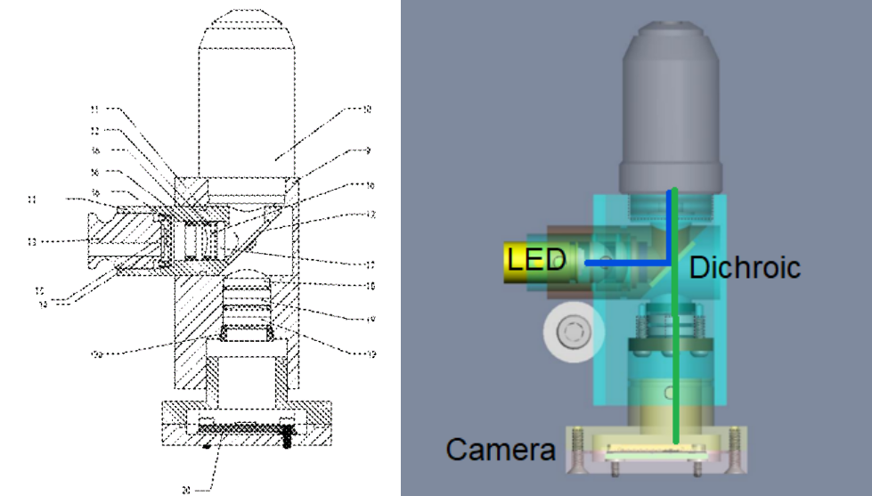
With advances in CMOS cameras, LEDs as excitation sources and multi-bandpass filter technology, Etaluma embarked on designing the simplest commercial incarnation of a high quality epi-fluorescence wide field microscope. The availability of USB CMOS cameras and their displacement of CCD designs allowed 5 VDC power over the high current requirements of a cooled/intensified CCD. LEDs are also driven at 5 VDC and so a single USB cable can power, control, and acquire images. A simple user interface allows image acquisition and time lapse automation (Figure 1).
Real Microscopy
Imaging small objects can be accomplished in many ways. Our goal was to preserve many of the features of conventional microscopy. A large and stable ecosystems of objectives, transmitted illumination, and c-mount camera standards were important to use when appropriate. Reducing the optics any smaller than the conventional scale results in lower sensitivity and the need to make one’s own objectives.
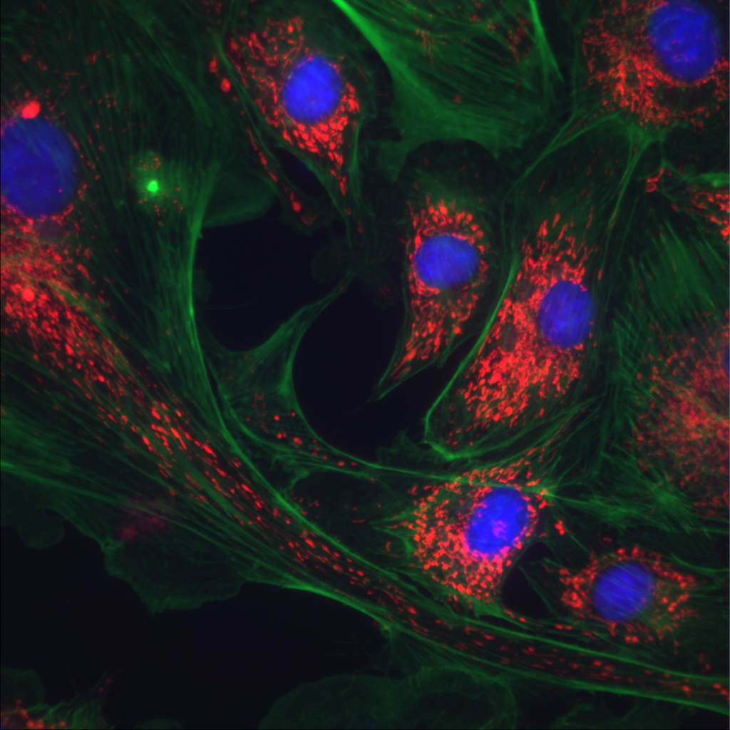
When designing a multichannel version, we chose a Pinkel filter set configuration. This multi-bandpass approach allowed the elimination of filter wheels and the associated mechanical complexity. The result was a dedicated three color capability with excitations for DAPI/Hoechst (405nm), FITC/GFP (488nm), and Texas Red and many RFP variants (590nm) (Figure 2).
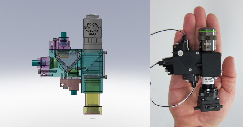
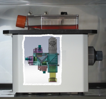
This optics module is mounted onto a focus mechanism, attached to a sample deck through a chassis with enclosure (Figure 3).
Killer Apps Emerge
Although the original goal was to simplify high quality microscopy, a few performance advantages emerged. By making the optics path as short as possible we preserved sensitivity. By eliminating as many optical elements (lenses, filters, and mirrors) we are able to limit losses and preserve the image quality delivered by the objective (the more elements in the path, the more critical the alignment and position of each).
The low power requirements led to an early customer placing the microscope inside their tissue culture incubator revealing live cell imaging and long term time-lapse as an important application. The high photonic efficiency of the design allowed much lower excitation energies to be used, dramatically reducing photo-damage from the long duration time lapse experiments. Measurements include simple proliferation, migration, fluorescent protein expression, and organoid monitoring (see user reviews). We have since had customers use the microscopes in 4 degrees C refrigerators and have tested operation to 90 degrees C (>200 deg F!).
The compact form factor made the design attractive to users who needed to add imaging to their existing instrumentation as well as new imaging based analysis instruments for research and the clinic. One application we could not have foreseen is microscopy in space. At the time of this writing, Etaluma has three microscope modules orbiting on the International Space Station. Obviously the size and weight of any payload is critical but the violence of the launch and the vibrational environment of the ISS is detrimental to conventional designs.
The Future
A double headed microscope, Figure 4 with two complete optics modules 36 mm apart is being used by our customers to expand the number of channels or double the imaging speed. Further advantages of “parallel” microscopy are just emerging. The most promising is its ability to cover larger field of views while maintaining high magnification. The time for tiling large FOVs is reduced by the number of microscopes simultaneously imaging. Highly multiplexed tissue imaging is a clear application.
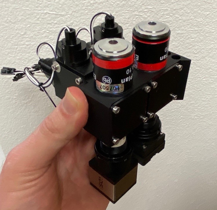
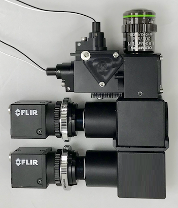
A wide variety of configurations and components have been supplied to our customers. Figure 5 shows a dual camera module for simultaneous 2 channel acquisition.
Things tend to get smaller, cheaper and more powerful; microscopy is no different. For more information visit our website at www.etaluma.com or to request someone contact you click here.
 (No Ratings Yet)
Write a Tools post
Create an account or log in to post your story on FocalPlane.
More posts like this

Filter by
- NewsApply
- DiscussionsApply
- How toApply
- ToolsApply
- Case studiesApply
- InterviewsApply
- JobsApply
- EducationApply
- Blog seriesApply
- Towards Global Acces..sApply
- Latin America Bioima..gingApply
- From Zero to Qupath ..HeroApply
- Asian Microscopists ..and Cell BiologistsApply
- AIC at HHMI JaneliaApply
- Deep Learning for Bi..o-image analysisApply
- GloBIAS – updates fr..om the communityApply
- WAMBIAN: West Africa.. in FocusApply
- Volume EMApply
- Latin American Micro..scopistsApply
- Bio-image Analysis w..ith NapariApply
- Imaging with…Apply
- Highlights from Euro..-BioImagingApply
- LSFM seriesApply
- DIY MicroscopyApply
- View all