Advertisement

vEM reveals the cellular composition of the Blood Nerve Barrier

Posted by , on 11 October 2024

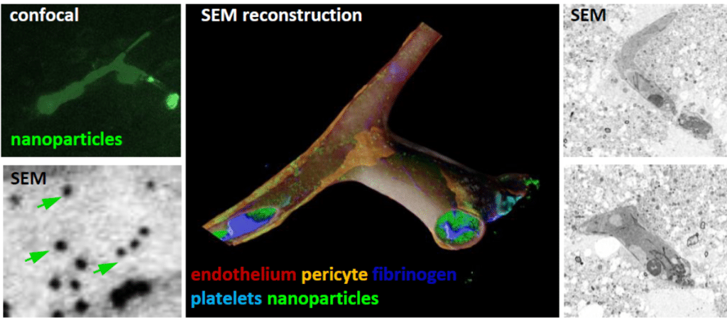
Using Serial Block-Face-SEM6 and AT-SEM, we characterized the cellular composition and interactions along EndoBVs7. Unlike the BBB, we observed that the vasculature within the endoneurium is not completely covered. Upon closer examination of the cells in contact with EndoBVs, we identified three distinct cell types frequently interacting with the vasculature.

Interview with Giulia Casal, the inaugural recipient of the Volume Electron Microscopy (vEM) Ambassador Travel Award!

Posted by , on 11 October 2024

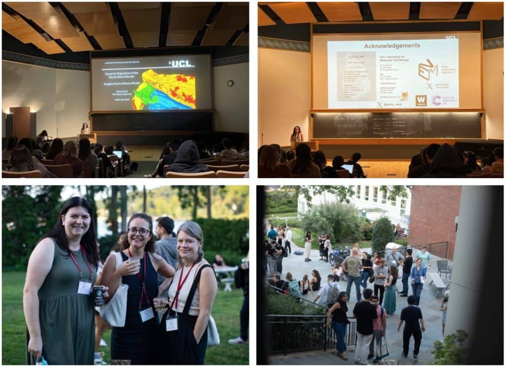
We congratulate Giulia for being the first recipient of the vEM Ambassador Travel Award! and thank her for advocating for our vEM Community! Giulia Casal is a PhD student in Prof. Alison Lloyd’s lab at the Laboratory for Molecular Cell Biology (LMCB) at University College London (UCL), where she is studying the blood nerve barrier

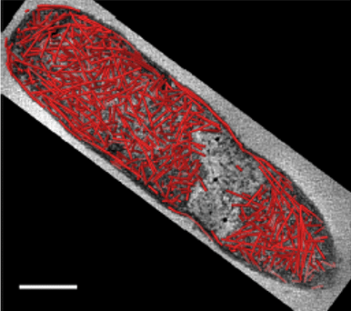
Kidney elastic fiber network changes in Marfan's Syndrome

Posted by , on 19 July 2024

by de Souza RB*, Meek KM#, Lewis PN#, Pereira LV*. *Department of Genetics and Evolutionary Biology, University of São Paulo, Brazil #StructuralbBiophysics Research Group, School of Optometry and Vision Sciences of the United Kingdom, Cardiff University, UK Challenge Marfan’s syndrome (MFS) is an autosomal dominant disorder that affects the connective tissue, resulting from a mutation

Targeted vCLEM of heterogeneous samples

Posted by , on 15 January 2024

by Karel Mocaer* and Paolo Ronchi# *Schwab Team and #EMCF, EMBL Heidelberg, Germany Challenge Studying cells or events within their biological context (i.e. in tissues, ecosystem etc…) is highly relevant. However, volume EM techniques are limited in the volume that can be acquired. Therefore, targeting approaches are needed to characterize specific regions of interest. This

Deciphering cardiac multiscale organization

Posted by , on 11 December 2023

by Sandra Rugonyi and Claudia López Oregon Health & Science University, Portland Oregon, USA Challenge The morphology, microstructure, and ultrastructure of heart tissues is exquisitely organized to optimize cardiac efficiency. In cases of cardiac malformations, known as congenital heart disease (CHD), spatially heterogeneous and abnormal ultrastructural patterns emerge as early as fetal stages that worsen

Insights into the leaky Blood Brain Barrier by vEM

Posted by , on 4 August 2023

by Martina Schifferer, German Center for Neurodegenerative Diseases, Munich Cluster for Systems Neurology SyNergy, Germany DOI: 10.1242/focalplane.15936 Challenge The Blood Brain Barrier (BBB) controls the exchange between blood and the brain parenchyme. Its structural determinants comprise tight junctions and endothelial vesicles which require resolution at the nanometer scale. In disease like stroke or traumatic brain

Inputs and Outputs of vEM in a Sensory System

Posted by , on 19 May 2023

by Federica Mangione, The Francis Crick Institute, UK DOI: 10.1242/focalplane.14809 Challenge The sense of touch enables organisms to interact with their environment by perceiving physical forces and guiding complex behaviors. Touch sensing is mediated by sensory neurons that innervate the body surface of animals and, together with surrounding cells, form specialized structures known as tactile

How volumeEM (vEM) can help map neuronal circuits

Posted by , on 2 March 2022

by Nadine Randel, University of Cambridge, UK DOI:10.5281/zenodo.6320414 Challenge The nervous system processes sensory information and generates different behavioural outputs accordingly. Synaptic resolution wiring diagrams form a basis for our understanding of how the brain works. Such a connectome provides the synaptic connectivity between all neurons and allows to identify all potential neuronal pathways which

Meet the people behind Volume EM community (part 2)

Posted by , on 28 December 2021

In this second post, we continue talking with some of the scientists involved in the Volume EM initiative. If you missed the first part of these series of interview, you can find it here. Raffa Carzaniga and Errin Johnson, Training Working Group What are the aims of your WG? The main aim of the vEM

Meet the people behind Volume EM community (part 1)

Posted by , on 21 December 2021

Volume Electron Microscopy or volume EM (vEM) is a relatively new term that brings together several recently developed imaging approaches that use scanning and transmission electron microscopy (SEM and TEM) to allow the interrogation of cell and tissue ultrastructure in 3D, at μm to mm volume scales and nm resolutions.​ In this blog series ‘volume