Turn Your Inverted Microscope into a Multimodal Nanoscope
Sponsored by Olympus, on 14 June 2021
Among recent nanoscopy techniques that break the diffraction limit, single-molecule localization microscopy (SMLM) contributes to major discoveries in medicine and biology. It is now possible to see how subcellular molecular machineries form and behave inside single cells and to quantify single biological molecules such as proteins, nucleic acids, and lipids, at ultralow concentrations inside the cell.
The crucial next step is to overcome these challenges that labs or core facilities face: understanding the complexity of the technology, educating researchers to new sample preparation protocols, and obtaining useful data from those acquisitions.
Abbelight has revolutionized the way of working in SMLM with innovative instruments and services adapted to all users:
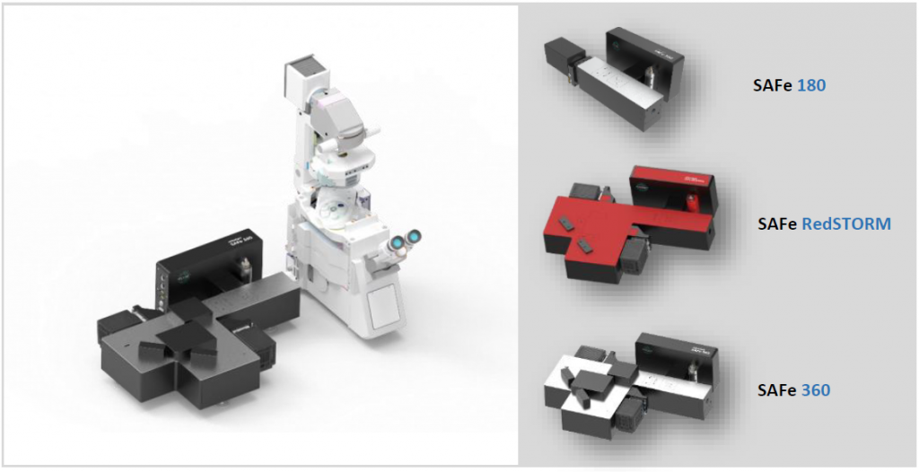
- Ground-breaking optical systems—The SAFe illumination device is an add-on module that can be easily integrated into any inverted microscope with a camera port. It transforms the microscope into a multimodal platform with advanced SMLM features.
- Specialized service—Bringing super-resolution microscopy within reach of both specialist and nonspecialist scientists, Abbelight provides dedicated, personalized support to users. It starts with deep study of your biological questions and close support throughout each step of your project through to data analysis.
Technical Solution
Abbelight’s technology enables any microscopist to easily access SMLM. The SAFe add-on modules range from the SAFe 180 (ideal for beginners), to the SAFe 360 (a multimodal imaging platform for advanced users), to the more recent SAFe RedSTORM (an innovative solution for multicolor spectral far-red demixing).
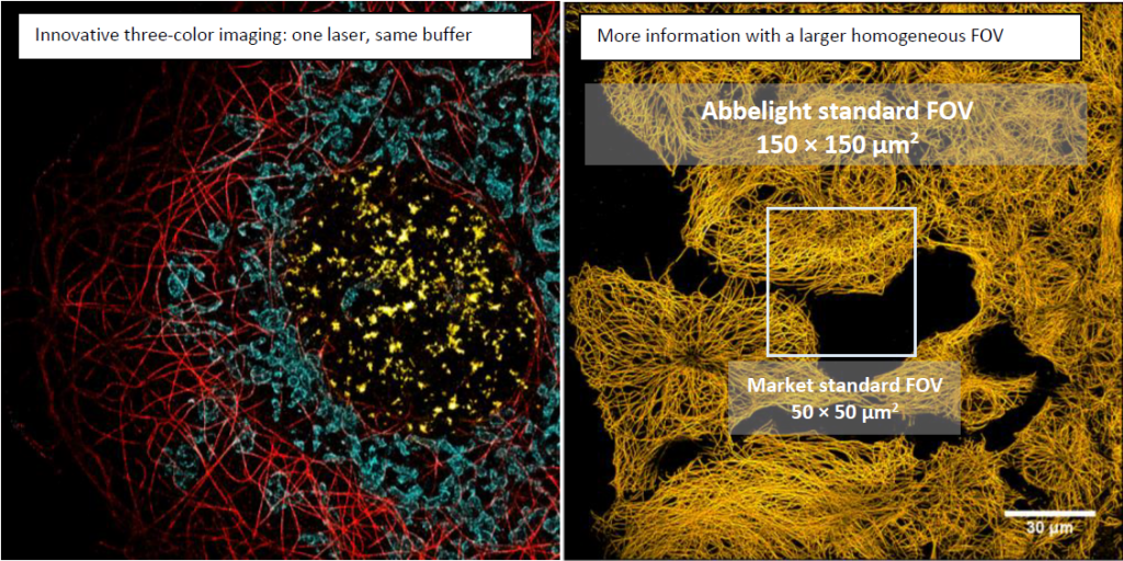
The modules all share the same unique excitation system, which is based on Adaptable Scanning for Tunable Excitation Regions (ASTER) technology.1 ASTER generates homogenous illumination in TIRF, HiLo, and Epi modes while performing SMLM modalities, such as PALM, STORM, or PAINT with a localization precision reaching 10–15 nm in 3D, on a FOV of 150 × 150 µm2.
| Excitation | FOV (obj. 100x) | Localization precision | 3D localization | Multi-color imaging | Spectral Demixing (SD) | SPT PALM | |
|---|---|---|---|---|---|---|---|
| SAFe 180 | EPI, HiLo, TIRF | 150 x 150 µm2 Homogeneous | < 15 nm | YES | Seq. | NO | YES |
| SAFe RedSTORM | EPI, HiLo, TIRF | 150 x 150 µm2 Homogeneous | < 15 nm | YES | Sim. | YES | NO |
| SAFe 360 | EPI, HiLo, TIRF | 150 x 150 µm2 Homogeneous | < 15 nm | YES | Seq. / Sim. | YES | YES |
ASTER Technology1 (Patent Pending):
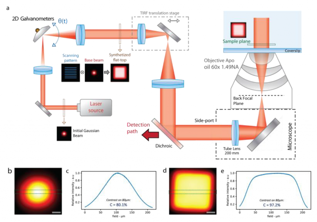
The ASTER illumination method is novel in its capability to exploit the entire FOV of sCMOS cameras for SMLM and TIRF imaging.
ASTER makes use of two galvanometer mirrors to control illumination at the sample plane. While the excitation beam keeps its position in the back focal plane (BFP), an angular rotation of a galvanometer induces a similar angle in the BFP, corresponding to a different position in the sample plane.
By applying specific patterns, such as raster scanning, ASTER can provide uniform excitation on tunable FOVs of up to 150 × 150 µm2 for all excitation modes (EPI, HiLo, and TIRF).
ASTER provides the flexibility that scientists seek, enabling them to adapt to various sample sizes and conditions. A prime example is when fluorescence density in SMLM is high or for particle tracking, the excitation area can be reduced to increase the irradiance or the acquisition speed up to 500 fps.
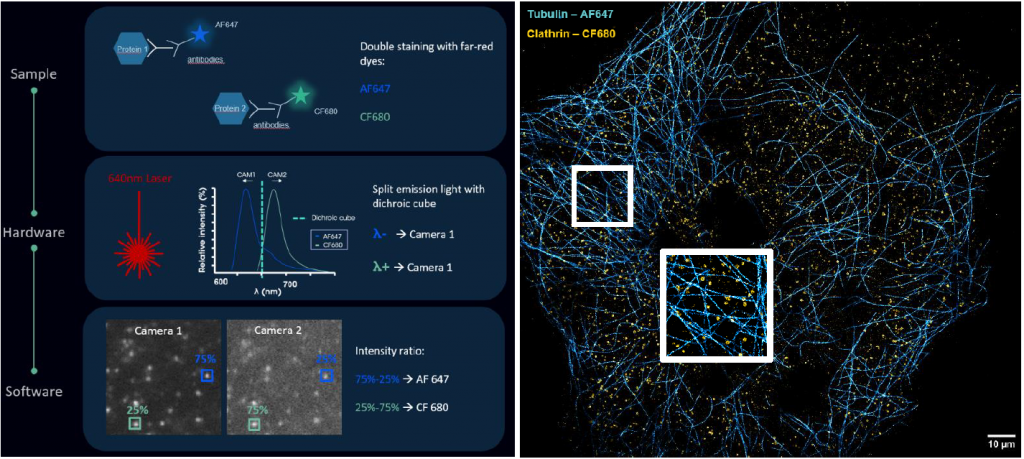
Multicolor Imaging by Spectral Far-Red Demixing
Although 3D nanoscopy has revolutionized the field of fluorescence microscopy by attaining unprecedented resolutions, multicolor imaging remains challenging in SMLM. This difficulty is due to several factors, including chromatic aberrations, the choice of optimal buffers, and the choice of single-molecule-compatible dyes.
To provide a solution, Abbelight has recently implemented spectral demixing for SMLM. By separating far-red dyes using a combination of the appropriate dichroic cube and ratiometric algorithms, this technique elegantly enables simultaneous multicolor imaging in SMLM. This advanced capability opens new avenues for researchers willing to push the boundaries of fluorescence nanoscopy. Spectral demixing is integrated in Abbelight’s dedicated instrument SAFe RedSTORM and SAFe 360 module.
In practical terms, it enables users to easily image in multicolor with one buffer, one laser, and one live acquisition.
Powerful Software for SMLM Data Analysis
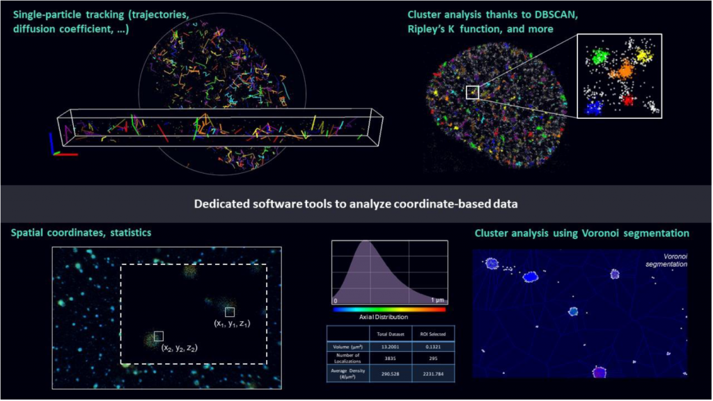
Unlike standard fluorescence microscopy, which generates pixel-based images, SMLM produces 2D/3D point clouds with millions of localizations and associated uncertainties. Thus, new computational methods are required for the quantification and interpretation of SMLM data to enable researchers to extract novel and groundbreaking biosignatures of biological structures and functions.
Abbelight’s NEO software offers powerful tools to study spatial and temporal distribution of localized single molecules, such as cluster analysis and single-particle tracking algorithms. Designed to be user-friendly and complete, NEO software simplifies data acquisition, provides real-time image reconstruction and quantitative feedback, and supports high-quality data and optimal SMLM data analysis.
Personalized Support
Abbelight provides a unique scientific support with SMLM expert scientists involved in multidisciplinary research, education, and international training. A dedicated expert is assigned to support the user in each step of SMLM projects: sample preparation, imaging, and data analysis. Users save time, acquire expertise, and have access to online tools on the Abbelight Academy (user guides, video tutorials, best practices, etc.).
A good experiment starts with good sample preparation, so ready-to-use and optimized SMLM kits are also available:
- Smart kit dedicated buffer for STORM imaging
- Blinking pad kit for non-adhesive cells for photo-activated localization microscopy (PALM) and STORM imaging
Collaboration of Innovators
Olympus and Abbelight recently joined forces to provide researchers with advanced and intuitive nanoscopy imaging systems. Abbelight’s vast expertise in SMLM and Olympus’ rich history in optical precision are the foundation of this collaboration. The SAFe modules can easily be incorporated into Olympus’ IX™ series microscopes to unlock the potential for SMLM microscopy. Olympus’ IX83 microscope is particularly well suited for nanoscopy applications due to its stable frame design, TruFocus™ Z-drift compensation system, and open structure, which facilitates multimodal operation. SAFe instruments have been successfully combined with Olympus’ FV3000 confocal laser scanning microscope as well as the IXplore™ SpinSR spinning disk system, enabling researchers to maximize their imaging capabilities with confocal, live cell super resolution, and SMLM in one system. Combined with Olympus’ industry-leading TIRF objectives, this collaboration delivers powerful and flexible nanoscopy imaging solutions.
For more information, contact your local Olympus sales representative at www.olympus-lifescience.com/en/contact-us/.
References
[1] A. Mau, K. Friedl, C. Leterrier, N. Bourg, and S. Lévêque-Fort. Fast scanned widefield scheme provides tunable and uniform illumination for optimized SMLM on large fields of view. Nature Communication. May 21, 2020.
 (4 votes, average: 1.00 out of 1)
Write a Tools post
Create an account or log in to post your story on FocalPlane.
More posts like this

Filter by
- NewsApply
- DiscussionsApply
- How toApply
- ToolsApply
- Case studiesApply
- InterviewsApply
- JobsApply
- EducationApply
- Blog seriesApply
- Towards Global Acces..sApply
- Latin America Bioima..gingApply
- From Zero to Qupath ..HeroApply
- Asian Microscopists ..and Cell BiologistsApply
- AIC at HHMI JaneliaApply
- Deep Learning for Bi..o-image analysisApply
- GloBIAS – updates fr..om the communityApply
- WAMBIAN: West Africa.. in FocusApply
- Volume EMApply
- Latin American Micro..scopistsApply
- Bio-image Analysis w..ith NapariApply
- Imaging with…Apply
- Highlights from Euro..-BioImagingApply
- LSFM seriesApply
- DIY MicroscopyApply
- View all