Meet the speakers: Hard X-ray Imaging of Biological Soft Tissues symposium (part 2)
Posted by Carles Bosch, on 6 October 2021
The Francis Crick Institute is hosting the 2nd edition of ‘Hard X-ray Imaging of Biological Soft Tissues symposium’ on Thursday 14 October 2021, 12:00-18:35 BST.
Here is the second post introducing some of the speakers participating in this symposium. You can read about the rest of speakers in our previous post. We hope you find some or all of these talks interesting and you join us on Thursday 14 October.
Marie-Christine Zdora “3D virtual histology through X-ray speckle-based phase tomography“
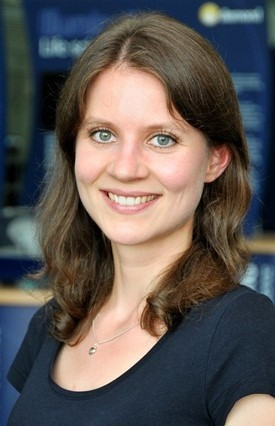
Marie-Christine Zdora completed a Bachelor of Physics (2011) and a Master of Physics (2014) at Technical University Munich, Germany, where she first started working on X-ray imaging. She also holds a Master of Medical Physics (2013) from the University of Sydney, Australia. For her PhD project on “X-ray phase-contrast imaging using near-field speckles”, she moved to Diamond Light Source, UK. In January 2020, Marie-Christine was awarded her PhD from University College London, UK, for which she received the Springer Thesis Prize. She then worked as a Research Fellow at the University of Southampton, UK, to develop her X-ray speckle-based imaging methods further. Since February 2021, she is a Postdoctoral Fellow at the Paul Scherrer Institut, Switzerland. To date, Marie-Christine has authored 38 papers and conference proceedings as well as a book. She has been an invited speaker at 21 international conferences, seminars and meetings in 8 different countries.
Talk description: X-ray speckle-based imaging (SBI) is one of them most recent X-ray phase-contrast imaging methods enabling the visualization of even minute density differences in a sample. It has received growing interest in the last years due to its simplicity, cost-effectiveness and robustness combined with the high phase sensitivity and compatibility with laboratory X-ray sources. Recently, SBI tomography has been identified as a promising candidate for virtual histology of biomedical tissue, providing 3D information at isotropic spatial resolution and quantitative information on the sample’s density. This talk will outline the principles of SBI and present our results on SBI phase tomography for 3D virtual histology of a murine kidney. Furthermore, a short outline of the possibilities and first results of SBI implemented at laboratory X-ray sources will be given, which is an important step towards the wider availability of the technique.
Ana Diaz “Ptychographic tomography for high-resolution X-ray imaging of biological soft tissues”
Ana Diaz has dedicated her entire career to develop X-ray characterization methods using synchrotron radiation. She did her PhD at the Paul Scherrer Institute in Switzerland on the characterization of colloidal suspensions confined in unidimensional gratings and then moved to the European Synchrotron Radiation Facility in France to work as a postdoc, where she used Bragg coherent diffraction imaging to characterize epitaxial SiGe nanocrystalline structures. She is now back at the Paul Scherrer Institute where she works as a beamline scientist at the Swiss Light Source since 2009. Ana has contributed significantly to the implementation of hard X-ray ptychography at the cSAXS beamline, in particular for ptychographic tomography. Together with her colleagues, she has received the Helmoltz Zentrum Berlin Innovation Award on Synchrotron Radiation in 2014 for high-resolution 3D hard X-ray microscopy.
Talk description: In this presentation I will first present the implementation of ptychographic tomography at the cSAXS beamline, explaining the current capabilities and showing the instruments that are available. I will then focus on the application of 3D imaging of soft biological tissues, detailing the sample preparation techniques that are possible and the current limitations in terms of contrast and resolution. Finally I will show a few examples of applications, e.g. in plant, retina, and brain tissues.
Carles Bosch “From circuits to synapses: systems neuroscience using hard x-rays (and light, and electrons)”
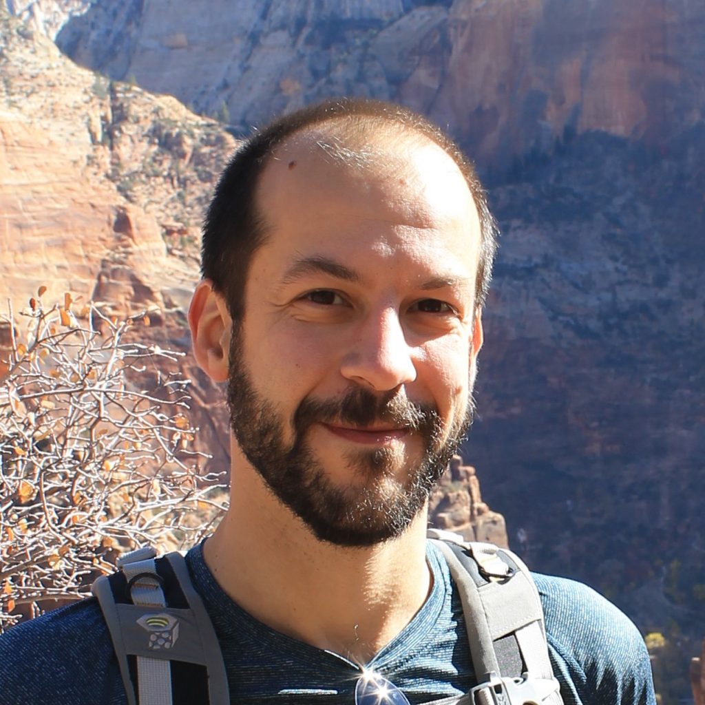
Carles Bosch is a neuroscientist at the Francis Crick Institute (London) specialising in correlative multimodal imaging of neural circuits. By combining an array of imaging techniques, he has developed a strategy to target specific regions in the brain to be explored with ultrastructural precision while keeping the big picture that provides context. He did his PhD at University of Barcelona, where he studied how synaptic plasticity is regulated in the adult mouse brain at the transcriptomic, molecular, and structural level. His current research addresses the structure and logic of the neural circuits for smell in the mammalian brain. Besides this, he is also interested in facilitating scientific discovery through multidisciplinary collaboration. He participates in diverse scientific outreach initiatives and has organised science-focused events tailored to both specialised and broad audiences.
Talk description: The brain is an organ that senses, computes and delivers information, which ultimately drives an individual’s behaviour. While the information flows through individual neurons, the computational logic is often embedded in neuronal circuits. In this talk I will compare techniques that employ light, X-rays and electrons and that can be used to image neuronal circuits in brain tissue. This will provide the context in which three particularly interesting hard X-ray imaging techniques operate: full-field, holographic and ptychographic tomography. Specific correlative multimodal imaging approaches will be discussed, showcasing how hard X-ray imaging is becoming an essential complement in the existing toolkit for systems neuroscience research.
Tim Salditt “Multi-scale 3d imaging of human brain tissue by X-ray holo-tomography”
Tim Salditt is professor of experimental physics at the Institute for X-ray Physics of the University of Göttingen. He studied physics in Munich and Grenoble, and received his Ph.D. in 1995 for research in kinetic growth of surfaces and interfaces, which he studied by diffuse X-ray scattering and non-specular reflectivity with Hans Peisl at the University of Munich. He then moved his focus to biophysics, using the newly developed approaches for interface-sensitive scattering to study fluctuations in oriented lipid membranes. In his postdoctoral work with Cyrus Safinya in Santa Barbara, he worked on the structure and interactions of lipid/DNA complexes. Driven by the motivation to study biomolecular assemblies also in the hierarchical and complex functional environment of cells, he developed X-ray waveguide optics for nanoscale holographic imaging and tomography.
With his group, he has designed a synchrotron radiation instrument, which they operate at the PETRAIII storage ring, together with DESY. Using a combination of diffraction with micro- and nano-focused beams as well as holographic imaging and tomography, they can now study biological matter, from the molecular level to biological cells and tissues. They push the limits in X-ray optics, including focusing, wave-front control, coherence, phase retrieval, reconstruction algorithms, information theory and image processing in order to get a maximum of information from a minimum of photons. Recently, they have got increasingly involved with implementing phase contrast phase contrast tomography for 3d mapping of the cyto-architecture, notably of brain tissue.
Tim Salditt has been spokesperson of the collaborative research center Nanoscale Photonic Imaging for 12 years, and is a principle investigator of the DFG Center for Excellence Multi-Scale Bioimaging: from molecular machines to networks of excitable cells. He is a member of the Academy of Science. In Göttingen, and of the DESY scientific advisory committee.
If Tim is out of Göttingen and not at a synchrotron beamtimes with his team, he may enjoy some family trip, and probably a mountain escape. Luckily, this can easily be combined at some synchrotron facilities.
Talk description: In my talk I will report on our recent work developing multi-scale phase contrast X-ray tomography (PC-CT) for 3d analysis of the cyto- and myolo-architecture of the human brain. This starts with continuous improvements in X-ray optics, nano-focusing, beam conditioning, combinations of parallel and cone beam geometry (Frohn et al., J.Synchr. Rad. 2020), phase retrieval (Lohse et al. J. Synchr.Rad 2020), image acquisition, and multi-scale analysis. In particular, I will discuss three challenges: First, how to extend the field of view towards entire brain regions, while preserving single cell and sub-cellular resolution, based on our recent multi-scale approach, as well as laboratory µCT (Toepperwien et al, Neuroimage). Second, quite on the contrary, how to reach higher resolution as required for fundamental connectomics. To this end, I will present a new data acquisition and inversion scheme, which we denote as super resolution X-ray holography (Soltau et al. Optica 2021). Third, how to use machine learning and concepts of optimum transport theory to exploit structural data for analysis of pathological changes associated with neurodegenerative diseases (Eckermann, Schmitzer, et al, umpublished). Finally, I will showcase present state-of-the art applications of PC-CT to human brain tissue
Meike Sievers “Large-scale connectomics with multibeam scanning electron microscopy”
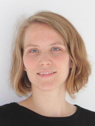
Meike Sievers studied physics at the University of Konstanz and the Ruprecht Karl University in Heidelberg. She first got in touch with Neuroscience during her 6-months Bachelor thesis at the Biology Department of the École normale supérieure in Paris. In 2014, she joined the group of Moritz Helmstaedter at the Max Planck Institute for Brain Research in Frankfurt am Main for her Master thesis project, where she mainly worked with serial block face electron microscopy. In 2016, she continued her work in the same group as a PhD student. Her PhD project aims scaling up dataset volumes for connectomic analysis using the Zeiss multi-beam electron microscope.
Talk description: My talk will be focused on the ATUM-MultiSEM workflow for connectomic data acquisition. I will discuss experimental and computational challenges of capturing and analyzing millimeter-scale volumes at nanometer-scale resolution, showing results from a petascale MultiSEM dataset from mouse somatosensory cortex covering all cortical layers.
Yannick Schwab “CLXEM & CXEM: when X-ray and fluorescence imaging lead the way to targeted volume EM.”
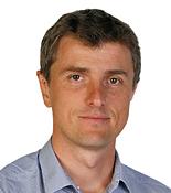
Yannick Schwab is a neuroscientist by training, graduating in 2001 at the University of Strasbourg, France. After two post-doctorates in Neurobiology (in Canada and in France), he joined in 2005 the staff of the Electron Microscopy facility at the IGBMC, Strasbourg, becoming its operational manager in 2009. During that time, he developed methods in Correlative Light and Electron Microscopy (CLEM) applied to cultured cells and to model organisms.
Yannick joined EMBL in 2012, as a team leader in the Cell Biology and Biophysics Unit and as head of the Electron Microscopy Core Facility (EMCF). The Schwab team is focused on methods development in volume CLEM, combining in particular fluorescence imaging of whole-mount specimens with volume EM. The EMCF offers access to a large portfolio of techniques in cellular EM, including ultrastructural analysis, 3D electron microscopy and CLEM on a variety of biological model systems.
Talk description: In this presentation, I will illustrate how fluorescence and X-ray imaging, with lab sources and at synchrotron beam lines, build powerful 3D maps of resin embedded specimens. With such knowledge of the sample, volume electron microscopy (in this case FIB-SEM imaging) can be efficiently targeted to one sub volume of interest. The combinations of such modalities, namely Correlative X-ray imaging and electron microscopy (CXEM) or correlative light and X-ray and EM (CLXEM) are efficient solutions to drastically increase the throughput of volume EM and opens to thorough biological investigations.
Darren Batey “Biological imaging with hard X-rays across the length scales”

Darren Batey is the beamline scientist of I13-1 coherence branch at the Diamond Light Source and has developed its user-friendly multiscale, multimodal, ptychographic endstation for use by the material, geological, and biological scientific communities. He has worked for many years on the development of ptychography in the visible, electron, and X-ray regimes and continues to push the boundaries of high-resolution imaging.
Talk description: Effective studies of biological systems require the combination of knowledge from across the length scales, linking localised properties with global behaviours. I13 is a multiscale and multimodal beamline consisting of two independent branchlines; targeting both the micron- and nano-scales, through direct- and reciprocal-space imaging, respectively. This presentation will give an overview of the beamline and discuss the latest results.
Alexandra Pacureanu “Deep and precise exploration of biological samples with hard X-ray nano-tomography and friends”
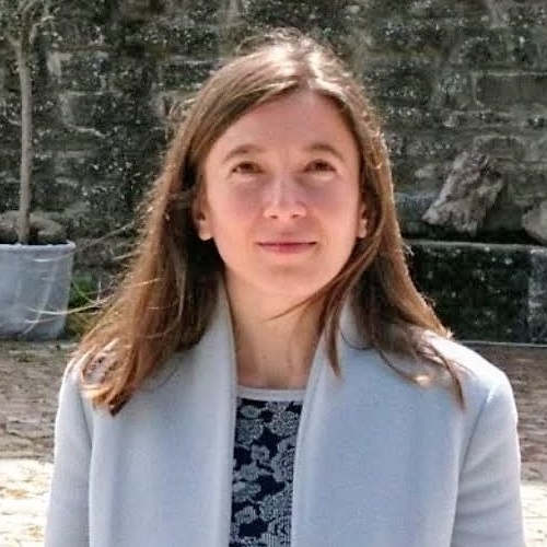
Alexandra Pacureanu is a scientist at the European Synchrotron leading the nanoscale X-ray neuroimaging unit. After completing her PhD in X-ray microscopy and image analysis at the Biomedical Imaging Centre, University of Lyon, and the European Synchrotron (ESRF) in France, she joined the Centre for Image Analysis at Uppsala University and the Science for Life Laboratory in Sweden for a postdoctoral fellowship. In 2014 she returned to the ESRF where, as a junior scientist, she focused on development of X-ray cryogenic nano-tomography for 3D imaging of biological tissues and cells. She continued her journey as a visiting scientist in the department of Neurobiology at Harvard Medical School in the US and senior research fellow in the Division of Biosciences at the University College London, UK before establishing her team at ESRF with support from the European Research Council.
Talk description: Recent advances in hard X-ray microscopy make possible reaching new levels of resolving power in thick biological samples. Thanks to high brilliance coherent beams combined with cutting-edge nano-focusing optics and high precision tomographic scanning, X-ray holography can probe the 3D structure of up to millimeter sized tissues at tens of nanometers spatial resolution. This opens new horizons for life sciences research, enabling to bridge scales and address questions previously unreachable. A prominent example is deciphering the structure and logic of neuronal circuits and their evolution with aging and diseases. This presentation will cover recent results obtained at the nano-imaging beamline ID16A of ESRF related to both fundamental life science questions and exploration of underpinnings and potential treatments for diseases. The ID16A setup is equipped with a cryogenic system, enabling imaging of vitrified samples and offering better preservation from radiation exposure. We will discuss the opportunities offered by hard X-ray microscopy as a standalone technology and in conjunction with complementary techniques, the current challenges to address, and perspectives in the context of the development of the fourth generation synchrotron sources.
Join the ‘Hard X-ray Imaging of Biological Soft Tissues symposium’ on Thursday 14 October 2021, 12:00-18:35 BST. Attendance is free. Register now.


 (No Ratings Yet)