An interview with Alenka Lovy
Posted by Mariana De Niz, on 27 November 2022
MiniBio: Dr. Alenka Lovy is an Assistant Professor at the Center for Integrative Biology (CIB) and in charge of LiSIUM, LightSheet Imaging at Universidad Mayor. where together with a team of researchers, she is introducing this novel platform to the Latin American region. Alenka is originally from the Czech Republic. She did her early studies in the USA, where she received her MSc degree in Plant Physiology studying Taxol biosynthesis in the lab of Dr. Rodney Croteau at Washington State University. She then did her PhD degree in Plant Biology in the lab of Dr. Peter Hepler at the University of Massachusetts. Then she moved to the Tufts School of Medicine in Boston, where she started working in a hybrid position as a Research Assistant Professor and managing the Tufts Imaging Facility. At Universidad Mayor she also holds a hybrid position, aiming to carry out her own research program on mitochondrial biology, and spearheading light sheet microscopy in the region.
What inspired you to become a scientist?
As a child I was interested in nature, but the inspiration to be a scientist didn’t quite come that early on. Trying to figure out the world around me and realizing you can understand how some parts of life and nature work led to my gravitation towards science. When the time came to choose a career path, I felt pursuing the understanding of life was the most honest way to find truth and be hopeful. I found this was the best way to spend my time, and I went for it. I liked the nobility of the search for knowledge. What I ended up studying was a BSc in Biology. I feel lucky I was able to follow a Biology major, and got the chance to work in several labs.
You have a career-long involvement in cell biology, metabolism, and microscopy. Can you tell us a bit about what inspired you to choose these paths?
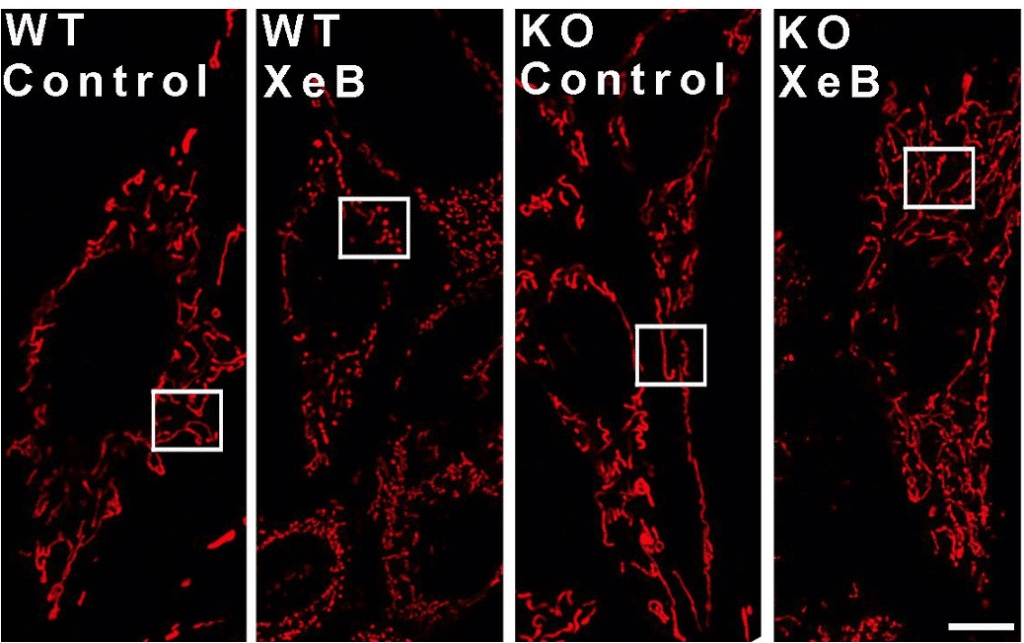
I was really interested in medicinal fungi when I started my career – but it was difficult to find mentors to help guide me on this path. I did find somebody out West studying Taxol biosynthesis, so I went to grad school to study biosynthesis of this anti-cancer compound and everything kind of evolved from there. Even though my path seems a bit disconnected, there are connections throughout it. After studying how this compound is formed inside the yew tree, I found it fascinating that these plants can produce very toxic chemicals and they themselves have to protect their own biology from these toxins. I learned about how compounds are sequestered in the cell, and how many of these chemicals are secreted out into the bark of the tree. I got really interested in the cell biology of that, and this is what eventually led me to microscopy. The compartmentalization of metabolites was really interesting to me, as well as understanding how they traffic within the cell. If one focuses on the cytoskeleton I feel there is so much light microscopy to do. I ended up dedicating my whole PhD to the study of the cytoskeleton. I first got my Master’s in Plant Physiology degree in Washington State University with Dr. Rodney Croteau where I studied Taxol biosynthesis part of the work. Then I moved to Massachusetts to study the actin cytoskeleton in pollen tubes with Peter Hepler who is a great microscopist. I was lucky that my advisor, who was actually called tubulin-Pete by his co-workers, was so experienced and passionate about microscopy. He was one of the founders of microtubules! His investigative spirit completely synchronized with mine, which led me to discover finer arrangements of actin seldom visualized before. It was here that I fell in love with microscopy and started helping in imaging facilities. I have been working in imaging facilities since then. First at the University of Massachusetts in Amherst and this led me to my job in Boston where I stayed for 10 years at a facility and trying to start my own research program. In Boston I collaborated on many projects and taught researchers to use the multiphoton, confocal, spinning disk and total internal reflection microscopes for their research. I began working much more with mitochondria in Orian Shirihai’s laboratory, and started developing mitochondrial assays within our microscopy core. Specifically, we were studying mitochondrial fusion, which I became very experienced at measuring with our multiphoton microscope. Together with Dr. Shirihai at Boston University I developed a faster high-resolution mt-PA-GFP screening assay to measure mitochondrial fusion using confocal microscopy. But being at a facility and working on my own project didn’t work out – it’s very difficult to do both, manage a facility and have your own research program. It’s too much for one person 🙂 It’s funny because now I’m in Chile doing a similar thing. At the same time it’s slightly different because here it’s all more focused. My main thing is the research, while the microscopy is more on the side. Here in Chile I work with Ramon Ramirez, who is dedicated to managing the microscopy side of things, and it’s very helpful. Working in the facility in Boston, I met a Chilean researcher, Cesar Cardenas, whom I started collaborating with. We investigated mitochondrial fragmentation during the inhibition of ER to mitochondrial calcium communication. We revealed that the presence of constant IP3R mediated calcium release taken up by the mitochondria coordinates its function with its morphology. In the absence of this Ca2+ signal, mitochondrial respiration decreases and eventually affects actin binding proteins that induce the fragmentation of the mitochondrial network. We propose that this particular mitochondrial Ca2+ uptake and the IP3R-MCU pathway are new therapeutic targets in cancer. More recently I participated in the characterization (Ca2+ signaling and bioenergetics) of a new IP3 receptor inhibitor. Eventually, Cesar and I got married, we have a child and we decided to move to Chile. We were looking for positions to work together, and the opportunity arose in Chile to have both of our labs together. We really want to do that. In the USA it was very difficult to find this opportunity, plus we wanted our daughter to also learn about Chilean culture and be in contact with that side of the family. Currently I am building a new research program investigating how commensal bacteria affect mitochondrial behavior during aging.
Can you tell us a bit about what you have found uniquely positive about working as a researcher in Chile and the differences and similarities you see between your education years in the USA and the education style you are involved in now?
I find that in Chile there is a very strong collaborative spirit which I’m amazed about. This collaborative spirit makes up for the lack of resources, and is something noticeably different from the USA. With having just only 3 years of experience here in Chile, most of which have been a bit sheltered, I notice a great respect for education at all levels, which is crucial to receiving a good education. It determines how far you can get. I feel the undergraduate studies here are more complete and students are better prepared for what the academic job is. Students at this level have experienced laboratory work, and get a good understanding of what it takes to do science. Because education is taken so seriously, students stay focused and can make progress. So despite it being harder to come across resources and having to do so much paperwork one becomes more organized and patient to get something done. This is a big difference because in the USA you get an idea and can implement it very quickly, whereas here in Chile it’s a bit slower.
Can you tell us a bit about your day-to-day work as a group leader and as a CZI grantee heavily involved in microscopy? Could you expand further on your strategy to expand global access to bioimaging?
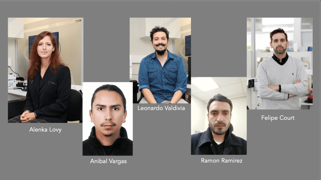
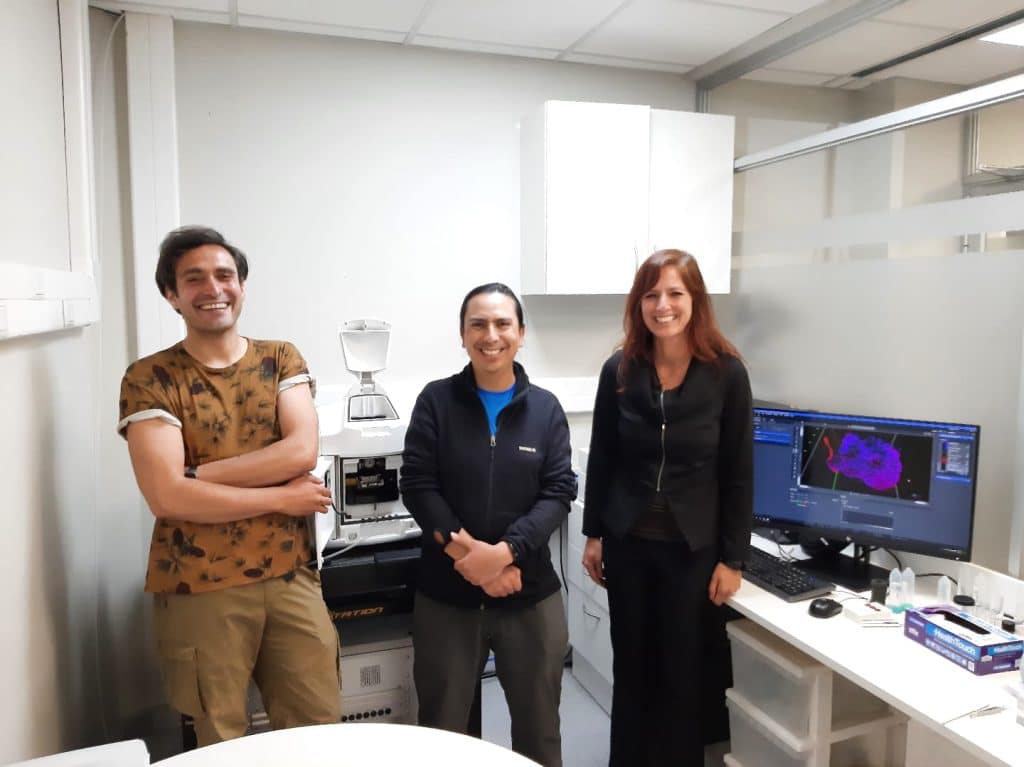
My laboratory is small. I have the help of a couple of research assistants and several undergraduate students each semester. Managing research projects, writing up research results into publications, applying for funding, organizing the lighsheet microscopy seminars and workshops, managing our collaboration in Germany to learn to build a lightsheet microscope and teaching courses keep me busy day to day. Slowly I am networking with microscopists in Chile, but also in Uruguay and in Brazil. I’m really excited about meeting light sheet researchers in South America. We’re trying to build a network through webinars, conferences and workshops but also we’re collaborating with Dr. Jan Huisken to learn to build economical light sheet microscopes which are portable. We reckon it would be brilliant to build these microscopes and be able to travel with them to show researchers across the whole country what you can do with them, to teach how they can be built if they are useful for those places. It’s something we’re working on. It’s a wonderful collaboration. Regarding the CZI grant, the director for the Centre for Integrated Biology, Felipe Court, forwarded me the call.Our university was just awarded with a ZEISS light sheet microscope, which was brought in by the other co-PI in this CZI grant, Leonardo Valdivia. Leo was successful with a FONDEQUIP grant to fund it the ZEISS light sheet. I believe it’s the only commercial equipment of this kind in the sub-continent, so we were thinking this is going to be a great way to share the instrument and try to build a light sheet bioimaging hub for the region. So we applied to the CZI grant and we were successful. These light sheets are so expensive, we realized not everyone is going to have resources to spread this technology. This is why I was very happy to know about the Flamingo light sheet and make the connection with Jan Huisken. He had already spoken to Leonel Malacrida about bringing the Flamingo to the continent and I am now trying to take the lead on this and am very excited about it. That is why it is so nice to be part of the Latin America BioImaging training working group, where we are gathering the training resources from the region.
Could you tell us more about your role in the LiSIUM initiative?
After successfully obtaining the CZI grant to start a light sheet imaging hub in Latin America, I have been leading the LiSIUM initiative organizing our website, seminars, workshops and conferences to bring light sheet researchers together to discuss advances in this imaging field. We are building our presence in courses, such as the EMBO Developmental Biology course taught annually in Quintay, the EMBO Global Exchange Lecture Course called Small Brains, Big Ideas, and we will also attend the annual Chilean Society for Cell Biology meeting. We hope to construct a mini-lightsheet attached to a cell phone in collaboration with Per Niklas Hedde and work with public schools to spread the lightsheet concept. I’m trying to find as many researchers that need light sheet as possible, so we can bring people together and share know-how on the techniques, protocols, etc. through webinars for example, and where we can solve questions together. Some people who already use confocals can really benefit from the light sheet and save a lot of time. You can image an entire organism in two minutes instead of spending hours and hours on the confocal. We’re also looking into expansion microscopy . As the resolution in the light sheet is not as high as in the confocal, some people could benefit from expanding their samples and using the light sheet. This is what our next webinar and workshop will focus on – the use of expansion microscopy with light sheet microscopy. We’re connecting with Steffen Härtel – they do very specialized light sheet techniques and are knowledgeable in image analysis, and I think it will be a nice collaboration. We also have remote operation where people can send their samples to us, but it’s a matter of advertising our work. I think through LABI and webinars and workshops, that’s what we want to achieve. So not everyone needs to travel to come and check the machine. They can just send the sample and still find out if it will be suitable for them. LiSIUM and the CZI project are very much hand in hand. Hopefully if we are all networked together, it will be easier for people to choose which technique is best for them. This is why spreading knowledge and access is vital.
Did you have many opportunities to interact with other Latin American groups, outside of Chile?
I have not had so much interaction with Latin American groups. During my PhD I worked with Luis Cardenas from Mexico and I think through him I was able to get some insight into the groups working in Mexico. But my biggest influence was Cesar Cardenas, who did introduce me to several Chilean researchers throughout the years we have been collaborating together. I was able to interact with some of his collaborators during my time in the USA. Other than this, I have not had much interaction. Hopefully this will start changing!
Who are your scientific role models (both Chilean and foreign)?
My mentors are my role models. Rodney Croteau, whom I did my MSc degree with, Peter Hepler, whom I got my PhD with, and Cesar Cardenas whom I have been collaborating with. I feel lucky that in addition to developing rich working relationships, I have developed meaningful friendships with my mentors. I value and respect this, and I aspire to be a good role model to others in the way my mentors have been to me. I learned a lot by observing how my mentors navigate the scientific field, I got to see their passion and perseverance. It’s been very inspiring and it aligns with my own expectations. Science is a hard and challenging field. I’m still learning how to navigate the Chilean system. But my mentors have taught me how to maintain that passion for science that helps you go through all the difficulties and troubles. I’m very appreciative of their contributions. I haven’t met so many scientists in Chile because of the pandemic, but hopefully I can find some role models here too. Right now I’m sticking to mentioning the ones I know well.
What is your opinion on gender balance in Chile, given current initiatives in the country to address this important issue. How has this impacted your career?
I feel Chile is quite progressive in the inclusion of women in the university setting. It was quite noticeable that the University has a gender equality initiative that they are trying to encourage. There could be more support by providing childcare at certain work functions to promote the building of community that would ultimately result in more scientific progress. Although there still seems to be some imbalance, there is a lot of initiative to try and close this gap.
Have you faced any challenges as a foreigner once you arrived in Chile?
The amount of paperwork is a challenge. It takes so much time that it’s hard to be efficient. Especially for a job that is so time-demanding, and then you add up all the paperwork. Additionally perhaps the language, but many people speak English. What I do sometimes is to talk with my students in English because many understand, but they respond back to me in Spanish, so we are all learning.
What is your favourite type of microscopy and why?
I think any live microscopy is my favourite, because I love being able to make movies of cells and then watching and hypothesizing what’s going on, trying to find explanations. I find this fascinating. It could be light sheet, confocal, differential interference contrast, DIC, anything that you can visualize a process with over time :). Watching ions changing during pollen tube growth – or seeing actin dynamics and wondering how it is coordinating tip extension. It’s super exciting trying to fit all the information you have from other experimental sources, and assembling it with the movie you just made.
What is the most extraordinary thing you have seen by microscopy? An eureka moment for you?
My eureka moment must have come when I was working with pollen tubes and I was looking for actin configurations in the apex of the cell. During live imaging we saw parallel actin bundles at the tip of the tube but we could not visualize them in fixed cells. So we couldn’t tell if what we were seeing was an artefact of injecting too much of the actin binding proteins to visualize the live actin cytoskeleton or if there was really some defined structure at the tip. My eureka moment came when I was able to prove what this structure was. What I did was to plunge freeze the pollen tubes in liquid propane to stop everything faster than chemical fixatives. When you freeze small samples in propane, its almost as if you vitrify or turn your sample into glass, because you can avoid ice crystal growth to damage the structure of your sample. Then you can permeabilize the cells to get antibodies into the different parts of the cell, and there was this beautiful sub-apical ring of actin with parallel bundles at the tip. It was an actin fringe in the cortex of the cell that we were able to visualize for the first time in fixed cells. It had been a controversial topic for many years and people weren’t sure whether this actin fringe existed because it did not show up in chemically fixed cells. Finding hard evidence to prove its existence was my contribution. I find it extraordinary to observing live pollen tube growth with DIC optics and seeing the fervent and almost stormy movement of vesicles at the point of cell extension, or watching the stages of mitosis and the extremely organized separation of chromosomes emerging from the chaotic and messy looking preparatory stages- it is breathtaking, and indescribable with words.
What is an important piece of advice you would give to future Chilean scientists? and especially those specializing as microscopists?
I feel like it’s a very good time now to become a microscopist. because there are many growing opportunities for this kind of future. Because there’s mechanisms in place to have someone evolve as a microscopist nowadays, I encourage it. Probably because I always wanted to be a microscopist and I had a pretty difficult time trying to maintain my position because of the lack of support and the lack of programs for this, I see this is changing. I built my own career and did my own trainings and found my own way, but a lot of it was quite unrecognized. I feel I spent a lot of time banging my head against the wall, not being able to network and exchange ideas with other scientists, and I’m very excited there are more avenues now in this direction. My biggest piece of advice is learn how to maintain your passion for science and your passion for knowledge because this is what keeps most scientists going through all the hard times.
Where do you see the future of science and microscopy heading over the next decade in Chile, and how do you hope to be part of this future?
This question is tough. I really hope the funding increases for science. The respect for science is already here, we just need more resources and funding so people can really bloom as scientists. I’m hopeful. With all the networking I feel we can make a difference. I see the CZI grant as a first step which has allowed us to organize webinars and training and has opened the possibility of bringing the Flamingo to South America. By networking imaging scientists together, we will be able to progress more efficiently. Personally, the CZI initiative has already made a huge difference because I have always worked from behind the scenes, supporting the research of others from imaging facilities while trying to run my own research program. However, now with the support of CZI and meeting many imaging scientists in a similar situation, our positions are becoming more recognized and integrated within the scientific field, hopefully creating more funding opportunities to keep our career paths growing.
Beyond science, what do you think makes Chile a special place to visit and go to as a scientist?
I love how there’s so many different regions that are unique in terms of food preparation, or medicinal plants for example.. I love traveling around here to learn all these differences. The fruits from the farms surrounding Santiago are amazing, as is the goat cheese from Cajon del Maipo, or Ulmo honey from the South of Chile, which is so unique and delicious. I look forward to visiting more of Chile now that we can. We live close to the mountains, so we love to go hiking. It’s impressive how many landscapes Chile can offer. You can travel to many beach towns across Chile’s enormous coast, and each one is unique. Just as a note, it’s very cold and very windy! I’m usually at the beach with a jacket, which is something new to me. But I get to watch the fishermen and see what they’re catching, I’d love to learn too!
 (2 votes, average: 1.00 out of 1)
Get involved
Create an account or log in to post your story on FocalPlane.
More posts like this

Filter by
- NewsApply
- DiscussionsApply
- How toApply
- ToolsApply
- Case studiesApply
- InterviewsApply
- JobsApply
- EducationApply
- Blog seriesApply
- Asian Microscopists ..and Cell BiologistsApply
- AIC at HHMI JaneliaApply
- Deep Learning for Bi..o-image analysisApply
- GloBIAS – updates fr..om the communityApply
- WAMBIAN: West Africa.. in FocusApply
- Volume EMApply
- Latin American Micro..scopistsApply
- Bio-image Analysis w..ith NapariApply
- Imaging with…Apply
- Towards Global Acces..sApply
- Latin America Bioima..gingApply
- From Zero to Qupath ..HeroApply
- Highlights from Euro..-BioImagingApply
- LSFM seriesApply
- DIY MicroscopyApply
- View all