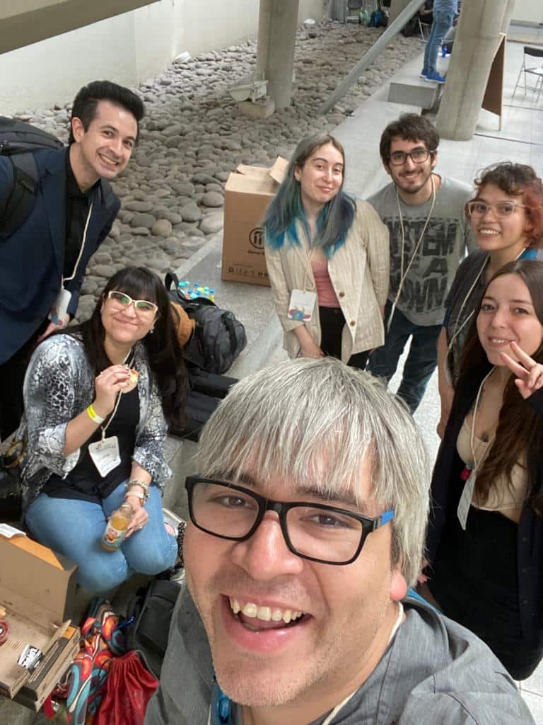
An interview with Jorge Toledo
Posted by Mariana De Niz, on 27 December 2022
MiniBio: Dr. Jorge Toledo is the director of the Advanced Microscopy Unit at Universidad de Chile. He has been heavily involved in multiple policies of scientific communication and outreach, as well as the democratization of microscopy and image analysis. He has spearheaded several initiatives both locally (in Chile) and regionally (in Latin America). Jorge did his BSc studies in Biochemistry at Universidad de Chile. In 2012 he began his PhD in the lab of Dr. Andres Couve focusing on organelle biochemistry, and it was here where he had his first approach to microscopy. He later did a postdoc in the labs of Steffen Härtel and Miguel Concha, where he began the establishment of what is now the Advanced Microscopy Unit which he leads.
What inspired you to become a scientist?
When I was a child, my school didn’t have enough practicals to get an idea of what science really way. Chemistry “experiments” were all about ordering orbitals in a piece of paper. I also didn’t know anything about microscopes at this time of my career. But around that time (in high school or so) we had some invited speakers to tell us about different career options. One of them said that being a Chemist was like being “architects of the cell”. They also conveyed the idea that science has no hierarchies – that scientists always discuss ideas as equals. Coming from a super hierarchical type of education where you mostly memorize things, the idea of being able to discuss ideas among equals was extremely appealing. I really wanted to become an “architect of the cell”, so this was my inspiration. I ended up studying a BSc in Biochemistry – the first two years are mostly Chemistry.
You have a career-long involvement in neuroscience, chemistry and microscopy. Can you tell us a bit about what inspired you to choose these paths?
I first worked on medicinal chemistry. At some point I found neuroscience appealing because Chile historically has a lot of involvement in this field, and we have the squid model too. So I joined a group working on neuroscience which also did a lot of microscopy. When I was doing my thesis I did not have a lot of access to the confocal microscope, because the user costs were about 25 USD per hour, and my salary was about 100 USD per month. It was unconceivable to use it. But at some point an Olympus microscope was brought to Chile for exhibition/testing – I had never seen a microscope as complex, but I was given the chance to learn for a whole week for free. I used it for a whole week, with very little idea of how to use it. But I ended up acquiring beautiful images and this was the start of my microscopy career. I did my PhD between 2012 and 2017 with Andres Couve Correa at Universidad de Chile. Last year andres Couvet became the Minister of Science in Chile. He gave me lots of opportunities to get involved in microscopy. Equally, during my PhD, I had a chance to spend a lot of time in Steffen Härtel’s lab, who is another expert in microscopy in Chile. With Andres Couve, we established STORM microscopy – we did this in collaboration with Daniel Choquet. Andres always supported me with all my interest and career as a microscopist. Later, between 2016 and 2019 I did a postdoc in two labs: with Steffen Härtel and Miguel Concha, also at Universidad de Chile. Here I started using microscopes in the context of Biophysics and Mechanobiology. But during my PhD and postdoc, we started applying to all the grants and funding opportunities relative to equipment, and every time we were awarded equipment grants that were related to my thesis or to my projects, I needed to learn more about these microscopes to be able to use them – I realized also that this was the perfect opportunity to get training for these microscopes – I would never had a chance to have such great training on a one-to-one basis. These microscopes we won grants for, included a lattice light sheet, a laser scanning confocal, a STORM and various others. While I was doing this, and since we had a lot of microscopes, the idea arose to create a facility, and in 2017 I was given the position of Director of the Advanced Microscopy Unit. This is the REDECA (Red de Equipamiento Cientifico Avanzado). This is now the current position I hold.
Can you tell us a bit about what you have found uniquely positive about becoming a researcher in Chile, from your education years?
Universidad de Chile organizes summer schools. They were very expensive and I couldn’t afford them, but I joined ‘incognito’ – these courses were integrative covering all subjects and you had a chance to join labs. I joined a neuroscience and physiology lab where you would receive small electric shocks to get the extremities to move 🙂 I barely understood what was going on – they were studies on nerve conductivity of course and it triggered my curiosity. I think these summer schools were fantastic – I found them very motivating to learn more and truly become a scientist.
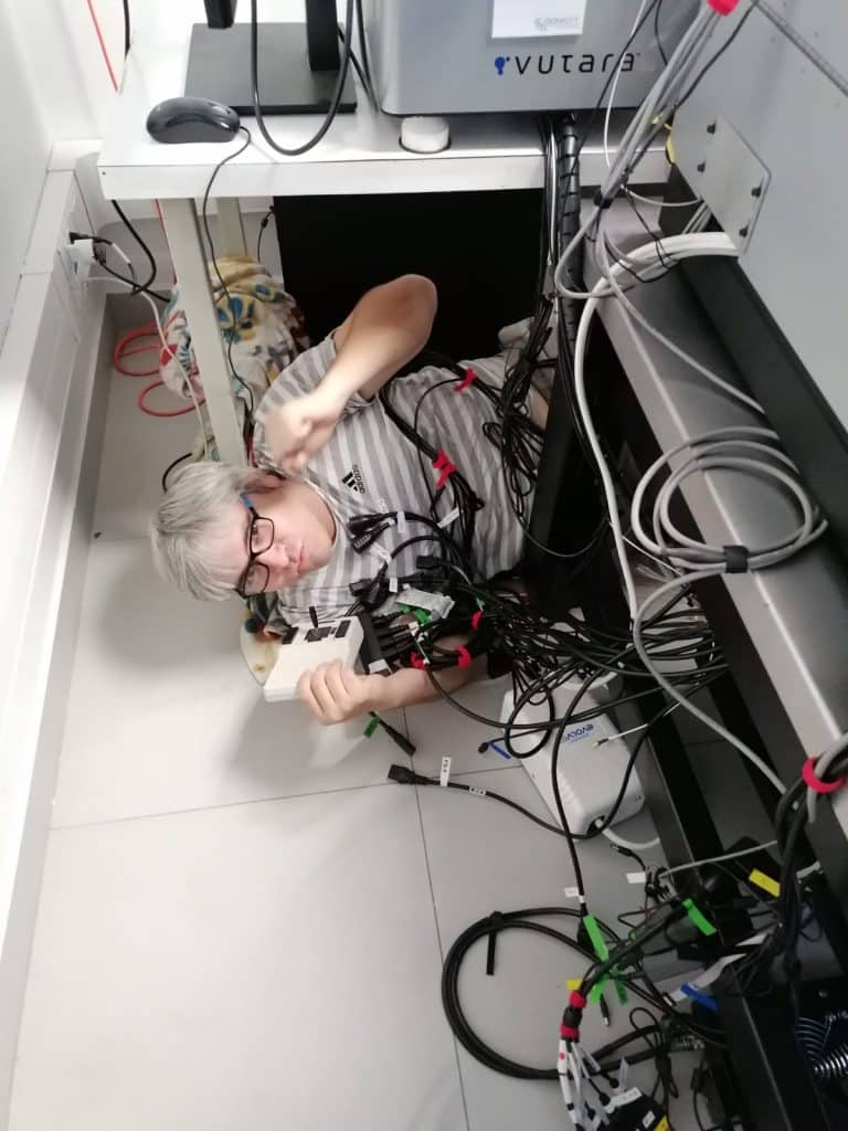
Can you tell us a bit about your day-to-day work as a director of the Advanced Microscopy Unit and REDECA?
During the pandemic, we brought together all the equipment of Universidad de Chile, so that they were all physically close. Now we’re optimizing everything and calibrating everything. I also have PhD students whom I can supervise. As part of their PhD thesis they all have to write a microscopy protocol, or a protocol for sample preparation. I feel they find this engaging – it becomes their ‘baby’. Depending on the time of the year, we might have additional stress: for instance, applying for equipment or research grants. I don’t think I have a routine, so it’s always entertaining and engaging. We also get engaged in a lot of different projects with different biological models, because of the teams that come to us seeking a service. In many cases, the service can go beyond that and become a collaboration. For instance, nowadays we have a collaboration investigating starch knives from Easter Island using FLIM; we are also investigating humming bird brains – we recently published a paper on a collaboration we did, studying mutations of the human brain. All these are collaborations – of course it depends on the main groups and how much of our involvement they want, but many times it’s very enjoyable.
Can you tell us a bit about your project MicroMundo?
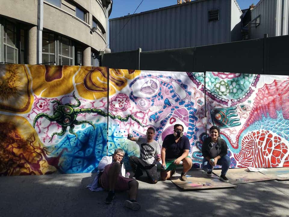
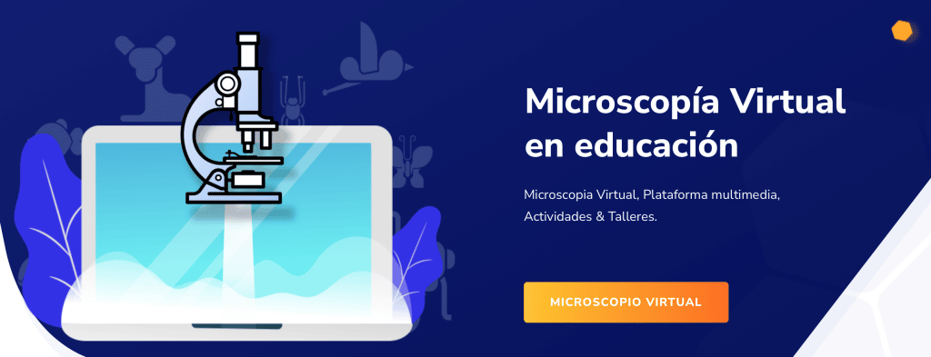
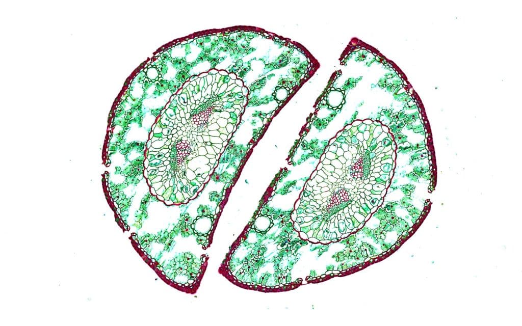
It arose because microscopes are expensive, difficult to handle, and sample preparation is difficult too. It requires significant expertise. Under this background, we wanted to ‘democratize microscopy’, and basically find ways to make it accessible to everyone through low-cost equipment. This included adapting the FoldScope and 3D printed microscopes. But since we have the tissue scanner which has platforms to make image digitalization on a large scale, we also took this as an approach to make image analysis accessible. First in workshops we asked students what they would like to see – their first answer was always blood. With a library of about 2000 images, we started doing other types of activities – to bring this to an educational context. Our latest ‘creation’ was to use ‘imaginary creatures’ (like Pokemons and Hello Kitty and characters of this sort) to teach young students how to perform CPR – we wanted them to learn about Anatomy: the names of bones, the position of organs, and so on. For example, we wanted them to learn about the relation between the heart, the brain and the lungs and how this is relevant to CPR. It was a very successful exercise, and the children became truly engaged. They would come later on and explain for example why Pikachu can run so fast, and how this might be related to the cerebellum and so on. It was helpful to allow them to make links between science and their favourite characters while having fun. I was impressed about this dynamic.
Eventually this was covered by the news! You can learn more about this initiative here: https://micromundo.team, https://www.uchile.cl/noticias/176855/conoce-el-proyecto-micromundo-en-el-capitulo-3-de-iniciativauchile Also, since in our University the microscope equipments are relatively under-utilized, we can use them for teaching and outreach purposes, and this also has been very successful. Our latest project with children was to determine how they perceive wine. It’s an agricultural school nearby, close to vineyards, and we take as a basis their knowledge on the soil pH and other factors important for wine, we then analyse together the stamps on the bottles to determine what everything means, and then we analyse the cultural background. We wrote a book about this during the COVID pandemic. It was a lot of fun. Other projects we have done include a short film about cells and planets, which we showed at the planetarium.
Can you tell us a bit about your involvement in SCEECA?
SCEECA is the Sociedad de Encargados de Equipamiento Avanzado. The idea was to bring together all the facilities in Chile to discuss common problems or situations: how to manage databases, booking systems, charging systems, conflict resolution with users if this arises, etc. All the management-related issues. We first made a national survey, and then organized a congress, where we analysed the results of the survey. One thing we realized is that at least in Chile, it’s uncommon that a facility manager is only dedicated to Imaging. Facilities are usually linked to Genomics, or Cytometry, or Pathology or other disciplines. Imaging is not considered a separate discipline. So our congresses bring us all together, and what we are doing now is legally formalizing the unit. We are waiting for the lawyers to finish this so we can apply to grants such as CZI as a unit. We also have a WhatsApp group where we communicate a lot to solve issues and discuss about Microscopy together.
Did you have many opportunities to interact with other Latin American groups, outside of Chile?
A bit. I had a lot of collaborations with the group of Federico Lecumberri (from Uruguay) because he came often to Chile. I also attended courses organized by Alfredo Caceres and Luis Bagatolli. Both were very open minded and they both introduced me to their network. They were very nice and really contributed to my career development. Also, I have many collaborators in Colombia – they have one of the largest deposits of brain tissue in the world. I love the country – it’s beautiful. So now I have about 4-5 collaborators there. And while I was there, I met other groups from Ecuador. We were recently awarded a grant of about half a million dollars to create a cloud with images for image analysis, which can be used by remote computing. There is one version in Uruguay, Peru, Ecuador, Colombia, and Chile. The aim is to have the whole map of Latin America with hubs: a LATAM network. In the latest LABI meeting I met Mexican scientists too, and that’s likely the next country we want to include in this project. Another thing we have been working on is a Biosafety and Biohazard map in the region, where we know where all the labs working with different pathogens in the region are located. So if someone wants to work with, say, V. Cholerae, they know which labs work on this and can go there to collaborate. But I must say all this expansion in Latin America is recent. Not so long ago, all our collaborators were mostly European or from the USA.
Adding to this point, I think there are many opportunities for collaborations and knowledge exchange in Latin America: for example, there’s Alianza del Pacífico, AUIP (Asociación Universitaria de Investigadores de Posgrado). I think it’s just about looking for these opportunities, because they do exist.
As a Chilean scientist, did you have any challenges working abroad?
I had a chance to work in Daniel Choquet’s lab in Bordeaux, France. I also went for short amount of time to Hamamatsu in Japan. I went there to learn how to use the tissue scanner. The rest has been mostly congresses and conferences. About differences, I noticed that in Japan everyone is very formal – lab meetings are not confrontational. People don’t criticize one another nor point out mistakes. I found this very frustrating because even if say, the boss was wrong, no one would point it out, out of politeness. I wasn’t there for a long time, but lab meetings were more of a conference than a lab meeting. In Bordeaux, the experience was different. For instance, since in Latin America resources are not plentiful, usually the same scientist that performs imaging, has to learn how to do image analysis. In Bordeaux, they had experts dedicated to each step of the microscopy pipeline, so I felt that sometimes people with different expertise didn’t make the effort to learn from the expertise of others. So during my time there I wanted to learn how to use a specific software, and nobody knew how to use it, except the one or two experts. When I asked those experts to teach me how to do it, they always offered to do it themselves. I really had to push, to be able to learn. I reckon the good thing in other countries of having so much specialized personnel is that you can publish much faster, but the downside is that you’re not forced to learn things. It’s the opposite in Latin American countries in my opinion.
Who are your scientific role models (both Chilean and foreign)?
I admire the way that Alfredo Caceres does science, always with a very critical mind. He develops things and collaborates with other people. Then he stopped a bit, took a sabbatical in Stefan Hell’s lab, then continued to learn things, then came back. As a PI he was very free-spirited and not within the general constrictions of academia. I feel many PIs sometimes take on many administrative roles and then become disconnected from what their lab members do. I also like a lot what Christian Wilson does and how he leads his lab. It’s a very small lab – only 6 people, and he is very hands-on. For me these two are role models.
What is your opinion on gender balance in Chile, given current initiatives in the country to address this important issue. How has this impacted your career?
I think there are important motivations to close this gap, but decision-makers in academia in Chile are still of a different generation who don’t see the importance of gender balance. I have heard still, at these high levels, misogynous comments and derogatory comments against women, including that they get positions just because they’re women (to cover a quota). I feel that the only way to fix this is issue is with time – when the generation that still has all these prejudices retires, the next generations will take over, with fresh ideas of gender balance. I still feel that currently many decisions to reach gender balance are “forced” and many people in power feel that many policies “affect men” and they only promote change because they feel obliged to do so, because it’s the trend everywhere else, rather than because they truly believe in this change. There’s no real consciousness. Many people in power here, still believe it’s a meritocracy, and that if women are not in high positions it’s because they haven’t worked hard enough, rather than acknowledging the glass ceiling and the gender differences. I think Chile still faces racism and sexism, and although it’s very prevalent, many people think it’s not real and it has no impact in people’s careers and lives. That’s my point of view! I’m currently in a group which aims to really push towards gender balance by putting pressure on issues where we realize gender balance is not being respected. It’s called ‘Comunica Ciencia Chile’.
What is your favourite type of microscopy and why?
That’s an excellent question. For my PhD thesis I used a lot the Yokogawa spinning disc microscope. I loved doing live imaging. I was studying organelles so live imaging was fascinating! I don’t have a lattice light sheet – perhaps if I had one this would be my favourite!
What is the most extraordinary thing you have seen by microscopy? An eureka moment for you?
Right now we’re implementing expansion microscopy, and one of my PhD students is establishing and optimizing these protocols to image different structures including the dendritic spines, and cilia. We were very surprised to see that the inside of cilia is relatively hollow. Also, since my PhD I have focused on organelles, and I can watch movies of the ER expanding and contracting for a long time and characterize the types of movement and other things. I love it.
What is an important piece of advice you would give to future Chilean scientists? and especially those specializing as microscopists?
On a practical level I would say, as a microscopist you should get all the trainings available. Attend workshops, go to courses, stay up to date all the time. Try to obtain and maintain access to people with excellent knowledge on microscopes. Moreover, image processing is a huge field and expanding much faster than microscopy itself. So try to keep up to date with this field too. When someone wants to work with me, I teach them how to segment 100 cells for a machine learning protocol so they know how it’s all done. Within microscopy labs, there are always new things to learn, and if you join one of them you’ll even learn things by osmosis. Another piece of advice is to start early and lose fear towards these huge machines. Sometimes students don’t want to touch them because they feel they’ll break them. This fear has to be eliminated.
Where do you see the future of science and microscopy heading over the next decade in Chile, and how do you hope to be part of this future?
This is a great question. I feel that after I give you my answer, I’ll keep thinking about it the rest of the day. I think super-resolution will become a huge field. But I think quickly the mathematics associated with images will overcome it. It will become more relevant when joined with machine learning. It will be lower cost, and will become a more dominant technique. I think also that the future of microscopy for a democratic approach might be the generation of centralized facilities, where anyone can come to do experiments without having to set up their own facilities if resources are limited. There will be less redundancy, and more open access and open development. Especially there should be a good accountability of the equipment that is already there, like what we did in REDECA – people can already look this up publicly and know what is available to the scientific community. I feel democratic science should be a philosophy to the life of all scientists. Maybe this will lead to more strategic planning of which equipment to purchase or which grants to pursue. Maybe then the focus will switch to doing more complex experiments and addressing novel research questions rather than replicating efforts within regions. With LATAM and maybe also LABI the purpose is to facilitate research: for example, it’s difficult to move biological samples around. So in centralized facilities across Latin America, if someone has a certain pathogen or plant or embryo or rare fish that other researchers want to use within the context of complex microscopy, they will be able to do this more easily. At present transporting these organisms is very complicated and expensive.
Beyond science, what do you think makes Chile a special place to visit and go to as a scientist?
Chile is now going through difficult times. Right now we had a big issue with our constitution. I feel sometimes Chile is very progressive but then things like the constitution issue happened, and it’s a setback. So it’s a difficult time right now. But Chile is a beautiful country- we have a connection with Antarctica, we have Tierra de Fuego, Easter Island and many other beautiful places. We have excellent fibre optics, which we can connect all the way from our major observatory to Antarctica. We’re a country of a lot of contrasts, which can be good and bad. At the moment with the rejection of the constitutional changes, it feels that this is what sometimes characterizes Chile: it’s stable in its way of thinking and in rejecting changes, which can be frustrating.
 (No Ratings Yet)
Get involved
Create an account or log in to post your story on FocalPlane.
More posts like this

Filter by
- NewsApply
- DiscussionsApply
- How toApply
- ToolsApply
- Case studiesApply
- InterviewsApply
- JobsApply
- EducationApply
- Blog seriesApply
- Latin America Bioima..gingApply
- From Zero to Qupath ..HeroApply
- Asian Microscopists ..and Cell BiologistsApply
- AIC at HHMI JaneliaApply
- Deep Learning for Bi..o-image analysisApply
- GloBIAS – updates fr..om the communityApply
- WAMBIAN: West Africa.. in FocusApply
- Volume EMApply
- Latin American Micro..scopistsApply
- Bio-image Analysis w..ith NapariApply
- Imaging with…Apply
- Towards Global Acces..sApply
- Highlights from Euro..-BioImagingApply
- LSFM seriesApply
- DIY MicroscopyApply
- View all