An interview with Cristina Bertocchi
Posted by Mariana De Niz, on 10 January 2023
MiniBio:
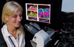
Dr. Bertocchi had a formal training in Biology during her undergraduate school in Italy at the University of Milan. She then moved to Innsbruck, Austria, where she embraced biophysics and obtained her Ph.D. from the Department of Physiology and Medical Physics (Medical University of Innsbruck, Austria) in2018. She then moved to Singapore for her postdoctoral training at the Mechanobiology Institute of Singapore. There she started to develop her interest in investigating molecular nanomachines regulating force transduction and mechanosensing. And the perspectives she developed there are essential to the research that she is currently leading in Santiago de Chile where she started her independent research career and where she is leading the laboratory for Molecular Mechanics of Cell Adhesion. She part of the UMA comitee (Advance Microscopy Unit) at Pontificia Universidad Catolica. She is Editorial Board Member of various International Journal. Since 1st of July 2022 she has been appointed Visiting Professors at University of Osaka.
What inspired you to become a scientist?
I have always been a very curious person, always asking why. At first, during my secondary school, I have been attracted to Genetics but then decided to move towards Physiology that explains how things work. There is a fantastic feeling that comes from understanding something, and unveiling how nature works. It brings excitement and every discovery gives you new pieces of a puzzle.
You have a career-long involvement in mechanobiology and microscopy. Can you tell us a bit about what inspired you to choose these paths?
When I first started to apply for a tenure track position I looked back at my academic career and tried to build a connection between everything I have been doing all over the years. In that moment I have realized that despite not knowing the name of the discipline (Mechanobiology), I had always been working with forces applied to biological systems, even before joining the Mechanobiology Institute in Singapore. Firstly, cell volume regulation, and then the biophysical properties of surfactant and surface tension. Always my best companion was microscopy. Indeed, I have been using microscopy since my very first step in a laboratory, for patch clamp experiments first, and it has been love at first sight. I probably felt the same kind of excitement like when von Leuwenhoek for the first time looked at microorganisms moving in a drop of water. And, I never let it go. By using more complex and advanced techniques, under the wing of Prof Tony Kanchanawong, I have learnt how to use microscopy to look into nanoscale structures and unveil the nanoscale architecture of cell adhesion complexes.
Can you tell us a bit about what you have found uniquely positive about becoming a researcher in Chile, from your education years?
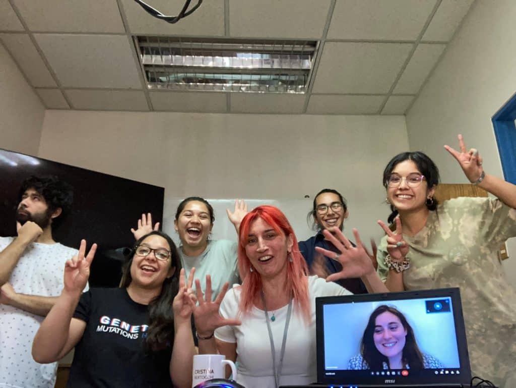
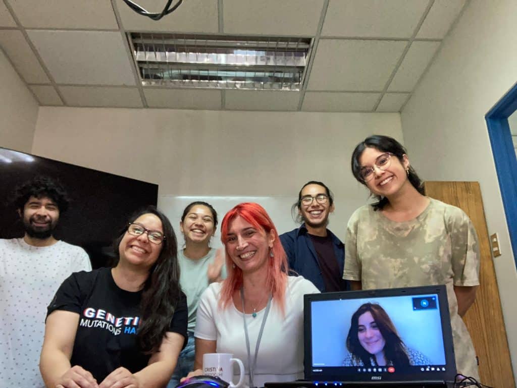
I came to Chile primarily because I was aware that there was little or no knowledge about mechanobiology. So, my husband (Dr. Andrea Ravasio, IIBM, PUC) and I decided to move to Chile with the desire to be the pioneers of this fantastic discipline in Latin America… and this paid off: we have two fantastic groups of students and collaborative projects that are running with success.
Can you tell us a bit about your day-to-day work as a group leader at the Pontificia Universidad Catolica de Chile, and as a member of the Advanced Microscopy Unit?
At Catolica we are lucky to have a good unit of microscopy (UMA) that allows us to do cutting-edge research. We indeed have the possibility to make use of microscopes such as the Airy scan, TIRF microscope, that, with minimal optimization can be used to perform superresolution microscopy. We have been recently awarded a big grant for the acquisition of Zeiss Elyra 7. Therefore, my day-to-day routine includes time at the microscope with members of my lab to do imaging and to teach them various techniques, and a lot of discussion with students and postdoc of the group about their results. On top of that, I do have quite a teaching load. My favorite subjects are obviously the Advanced microscopy course and the Physical bases of Biological processes, as there I feel I can really contribute to the education of the young researchers of the future.
Did you have many opportunities to interact with other Latin American groups, outside of Chile?
I am member of the latambioimaging, which provides a platform for the bioimaging community across Latin America with the aim of interact, collaborate and facilitate access to bioimaging resources. Furthermore, I am part of the QUAREP, a large group of scientists, facility managers, editors and microscopy company people with interest in improving quality assessment and quality control in light microscopy (https://quarep.org/). This association set me in contact not only with scientists from Latin America, but from all around the world.
Who are your scientific role models (both Chilean and foreign)?
Dr. Clare Waterman (NIH Distinguished Investigator) with her Nature paper in 2010, together with my former mentor at MBI (Tony Kanchanawong) and other colleagues, took advantage of three-dimensional super-resolution fluorescence microscopy to describe for the first time, at the ultrastructural level, the blueprint for how proteins in focal adhesions are organized. They then used this blueprint to explore the relationships between molecular perturbations, physical relationships, and force capabilities. Interestingly, I have applied similar approaches to study cell-cell adhesion complexes.
What is your opinion on gender balance in Chile, given current initiatives in the country to address this important issue. How has this impacted your career?
Despite all the progress Despite all the progress made and all the ground conquered, there is still quite an evident gender gap in science and research in Chile. In the department of Physiology, where I work, for example, there is an obvious imbalance in the number of female researchers (currently we are 2 from a total of 11 professors). Studies found that between 2015 and 2019 only 30% of new hires were women: indicating a clear gap. On top of this, a very low percentage of women leads national grants (individual or team related). So, recently, there has been an effort represented by the InES Gender Project that aims to help to raise awareness of gender inequalities by making the existing gaps in the production of knowledge and in academic careers more visible. This will hopefully open up to more discussion about the topic.
What is your favourite type of microscopy and why?
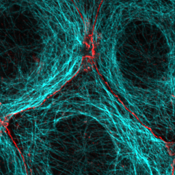
TIRF (Total Internal Reflection Fluorescence) microscopy because it is a very elegant technique, that allows imaging of fluorescent molecules located close to the interface eliminating the background. So, by reducing out of focus light, it can effectively increase the resolution of the system, that is the capability of differentiating structures that are separated in reality. The beauty of this system is that by simple optimization it can be used to achieve superresolution microscopy to image nanoscale details. This allowed us to create the blueprint of the cadherin-mediated adhesions (adhesive contacts between cells) with nanoscale precision (Bertocchi et al; Nat Cell Biol 2017). We are currently using it for SAIM (scanning angle interference microscopy) and adapting it for PALM and STORM.
What is the most extraordinary thing you have seen by microscopy? An eureka moment for you?
I mapped the nanoscale architecture of the cell-cell adhesion complexes. Eureka moment though has been when I saw for the first-time alveolar type II cells releasing surfactant. So exciting to think that what I was looking under the microscope, is something that constantly happens in our alveoli. At every breath….Thanks to microscopy we mapped the nanoscale architecture of the cell-cell adhesion complexes, and unraveled the molecular mechanisms regulating force transmission therein with previously unattainable details. This study posed the basis for further assessment of mechanistic details regulating cell-cell adhesion in physiology and disease, which is the main interest of our laboratory. Nevertheless, that Eureka moment for me has been when, during my PhD, I saw for the first-time alveolar type II cells releasing surfactant. So exciting to think that what I was looking under the microscope, is something that constantly happens in our alveoli at every breath.
What is an important piece of advice you would give to future Chilean scientists? and especially those specializing as microscopists?
As scientists, we learn that research starts with observation of ¨something¨ you are interested in to then form a hypothesis that should be tested experimentally. So, open your eyes and see through the lenses of a microscope and observe, observe observe: you can learn a lot simply by observing. And then, analyze your images!
Where do you see the future of science and microscopy heading over the next decade in Chile, and how do you hope to be part of this future?
I think the job of us microscopists is to spread the word. We should teach our colleague scientists what microscopy can do and how powerful it is. Then, researchers in Chile will have great opportunities like the most well recognized institutions worldwide.
 (1 votes, average: 1.00 out of 1)
Get involved
Create an account or log in to post your story on FocalPlane.
More posts like this

Filter by
- NewsApply
- DiscussionsApply
- How toApply
- ToolsApply
- Case studiesApply
- InterviewsApply
- JobsApply
- EducationApply
- Blog seriesApply
- Volume EMApply
- Latin American Micro..scopistsApply
- Bio-image Analysis w..ith NapariApply
- Imaging with…Apply
- Towards Global Acces..sApply
- Latin America Bioima..gingApply
- From Zero to Qupath ..HeroApply
- Asian Microscopists ..and Cell BiologistsApply
- AIC at HHMI JaneliaApply
- Deep Learning for Bi..o-image analysisApply
- GloBIAS – updates fr..om the communityApply
- WAMBIAN: West Africa.. in FocusApply
- Highlights from Euro..-BioImagingApply
- LSFM seriesApply
- DIY MicroscopyApply
- View all