Insights into the leaky Blood Brain Barrier by vEM
Posted by Georgina Fletcher, on 4 August 2023
by Martina Schifferer, German Center for Neurodegenerative Diseases, Munich Cluster for Systems Neurology SyNergy, Germany
DOI: 10.1242/focalplane.15936
Challenge
The Blood Brain Barrier (BBB) controls the exchange between blood and the brain parenchyme. Its structural determinants comprise tight junctions and endothelial vesicles which require resolution at the nanometer scale. In disease like stroke or traumatic brain injury the BBB is locally disrupted. The morphological hallmarks of BBB leakiness are,
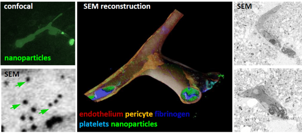
however, poorly understood. In our study1 we aimed at the identification of sites of BBB disruption in a mouse model of traumatic brain injury. These spots were visualized at the ultrastructural level by correlated light and volume electron microscopy (vEM).
Technique: ATUM CLEM
The idea of tissue slicing has been carried over from the Greek word ‘anatomy’ (‘through’ ‘cutting’) to modern volume electron microscopy (vEM). Among them, array tomography (AT)3 approaches are based on serial ultramicrotomy, collection onto solid support and serial scanning EM. The AT method automated tape-collecting ultramicrotomy (ATUM) was initially developed for large-scale neural circuit reconstruction4 while vEM of more
confined volumes has broadened investigation of a wide range of biological systems. ATUM enables the creation of a tissue section library that can be inspected by scanning electron microscopy (SEM) at different resolution regimes. Hierarchical imaging and the modular character of ATUM provide flexibility. Therefore, ATUM is not only valuable for the volume rendering of biological structures at the nanoscale but provides beneficial features for correlative light and electron microscopy (CLEM). We used 30 nm sized fluorescent and electron-dense nanoparticles to localize vessels of leaky BBB1. These target sites were relocated in the ultrastructural volume, generated by ATUM-SEM1. We observed nanoparticles within pericytes and abluminally1. Moreover, the ultrastructural volume captures the entire tissue context including all cells involved in an unbiased fashion.
Impact
- CLEM approach to target sites of leaky BBB
- Visualization of ultrastructural features of BBB disintegration
- Analysis of target sites for potential pharmacological intervention
References
1Khalin et al., Small, 2022; 2Peddie et al., Nat Rev Methods Primer; 3Micheva and Smith, Neuron, 2007; 4Kasthuri et al., Cell, 2015
 (1 votes, average: 1.00 out of 1)
Get involved
Create an account or log in to post your story on FocalPlane.
More posts like this


Filter by
- NewsApply
- DiscussionsApply
- How toApply
- ToolsApply
- Case studiesApply
- InterviewsApply
- JobsApply
- EducationApply
- Blog seriesApply
- Latin American Micro..scopistsApply
- Bio-image Analysis w..ith NapariApply
- Imaging with…Apply
- Towards Global Acces..sApply
- Latin America Bioima..gingApply
- From Zero to Qupath ..HeroApply
- Asian Microscopists ..and Cell BiologistsApply
- AIC at HHMI JaneliaApply
- Deep Learning for Bi..o-image analysisApply
- GloBIAS – updates fr..om the communityApply
- WAMBIAN: West Africa.. in FocusApply
- Volume EMApply
- Highlights from Euro..-BioImagingApply
- LSFM seriesApply
- DIY MicroscopyApply
- View all