Deciphering cardiac multiscale organization
Posted by Georgina Fletcher, on 11 December 2023
by Sandra Rugonyi and Claudia López
Oregon Health & Science University, Portland Oregon, USA
Challenge
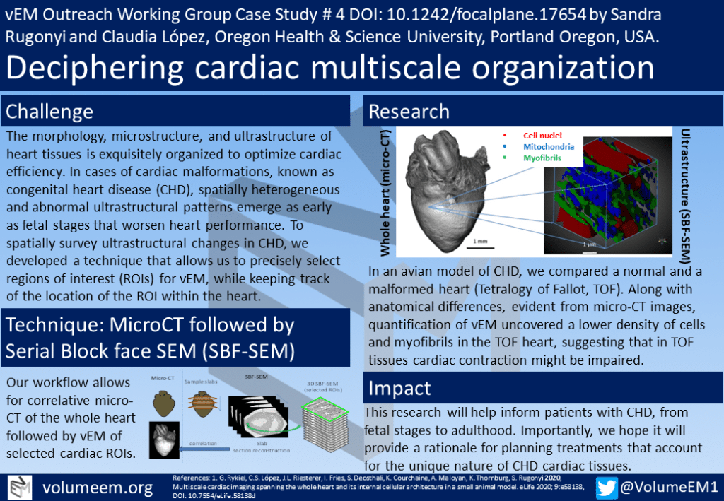
The morphology, microstructure, and ultrastructure of heart tissues is exquisitely organized to optimize cardiac efficiency. In cases of cardiac malformations, known as congenital heart disease (CHD), spatially heterogeneous and abnormal ultrastructural patterns emerge as early as fetal stages that worsen heart performance. To spatially survey ultrastructural changes in CHD, we developed a technique that allows us to precisely select regions of interest (ROIs) for vEM, while keeping track of the location of the ROI within the heart.
Technique: MicroCT followed by Serial Block face SEM (SBF-SEM)
Our workflow allows for correlative micro-CT of the whole heart followed by vEM of selected cardiac ROIs.

Research
In an avian model of CHD, we compared a normal and a malformed heart (Tetralogy of Fallot, TOF). Along with anatomical differences, evident from micro-CT images, quantification of vEM uncovered a lower density of cells and myofibrils in the TOF heart, suggesting that in TOF tissues cardiac contraction might be impaired.

Impact
This research will help inform patients with CHD, from fetal stages to adulthood. Importantly, we hope it will provide a rationale for planning treatments that account for the unique nature of CHD cardiac tissues.
References
1. G. Rykiel, C.S. López, J.L. Riesterer, I. Fries, S. Deosthali, K. Courchaine, A. Maloyan, K. Thornburg, S. Rugonyi 2020, Multiscale cardiac imaging spanning the whole heart and its internal cellular architecture in a small animal model. eLife 2020; 9:e58138, DOI: 10.7554/eLife.58138d


 (No Ratings Yet)
