An Interview with Dr. Harikrushnan Balasubramanian
Posted by Subhajit Dutta, on 2 October 2024
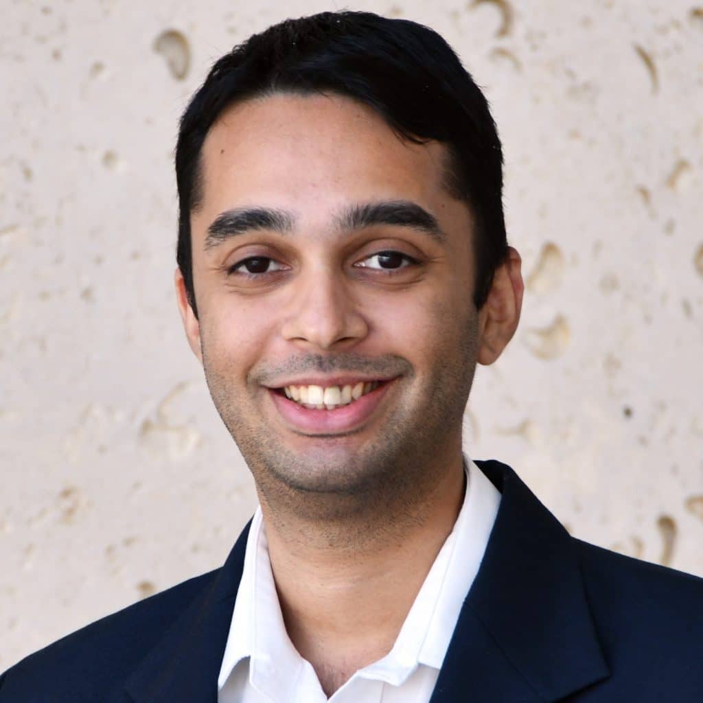
Welcome to the FocalPlane Interview Series where we spotlight groundbreaking Asian-origin scientists pushing the boundaries of cell biology and microscopy. Today, we are thrilled to present Dr. Harikrushnan Balasubramanian, a distinguished research specialist at Janelia’s light microscopy core facility. In this engaging interview, he discusses his path to a career in science, his specialization in fluorescence microscopy, and his dedication to fostering collaboration and training opportunities in underserved regions. Join us as we explore the exciting developments and future prospects in microscopy and cell biology through the lens of Dr. Balasubramanian’s expertise.
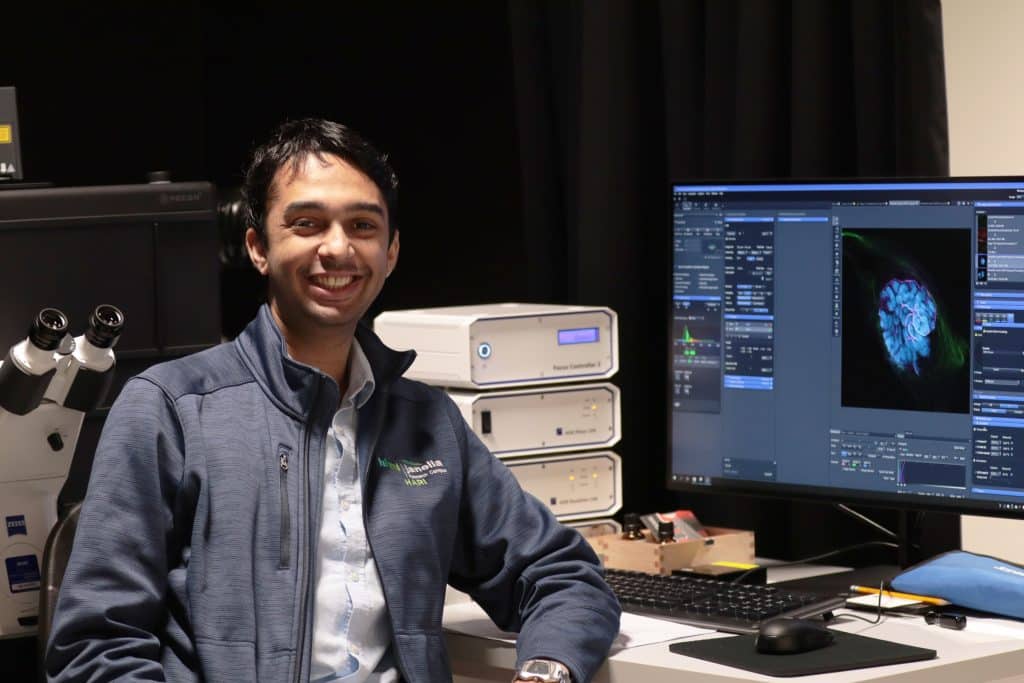
Bio in Brief: Dr. Harikrushnan Balasubramanian hails from Chennai in India. He obtained a bachelor’s degree from SRM University (India), followed by a PhD from the National University of Singapore (Singapore). After completing his doctorate, he joined the Advanced Imaging Center (AIC) at the Janelia Research Campus of the Howard Hughes Medical Institute (USA) as an Advanced Microscopy Fellow. He is now a Research Specialist in Janelia’s light microscopy core facility.
Q: Could you share your journey into science? What sparked your interest in cell biology and microscopy?
A: My passion for science wasn’t ignited by a single event, but rather a confluence of influences. As a child, I immersed myself in science fiction novels (especially Isaac Asimov) and the ‘Young Discoverer’ book series, fascinated by the wonders of the natural world – ranging from the cosmos to the microscopic realm. Movies like ‘Jurassic Park’ and even cartoons like ‘Pokémon’ further fuelled my curiosity about biology.
When it came to choosing an undergraduate path, I recognized that I was drawn more towards biology rather than mathematics-heavy subjects. In India, there’s a prevalent view that medicine is the primary career path for biologists. Since I wasn’t interested in that route, the best option was to pursue a degree in biotechnology.
Microscopy, however, didn’t become a major focus until later in my academic journey. During my undergraduate years, it was merely one of many tools used in lab sessions, where we observed cells or bacteria under a compound microscope. It wasn’t until my PhD that I delved deeper into the world of microscopy and appreciated its immense potential as a tool for discovery.
Q: What led you to pursue your PhD at the National University of Singapore (NUS)?
A: After completing my undergraduate degree, I was resolute in my desire to continue in science. Knowing that a PhD is often essential for many scientific careers, I chose to apply directly to a PhD program and save the additional time needed for a master’s degree. At that time, direct PhD opportunities in India were limited, which prompted me to explore options abroad.
I applied to several institutions in the US, Europe, and Singapore. NUS offered me a place in their biological sciences PhD program. This opportunity particularly appealed to me because, apart from the fact that NUS is a respected institution and boasts a vibrant and diverse international research community, Singapore is close to home, and is a safe and convenient city.
Q: What were your key observations and takeaways from your PhD experience?
A: I completed my PhD under Prof. Thorsten Wohland in an interdisciplinary lab focused on the biological applications of fluorescence correlation spectroscopy (FCS), a technique for studying molecular diffusion. My research concentrated on the epidermal growth factor receptor, where I explored how cell membrane properties affected its diffusion, clustering, and interactions.
My PhD experience at NUS was transformative in several ways. The international research environment provided invaluable opportunities for collaboration and exposure to diverse cultures, greatly enriching my learning experience. The ample funding, streamlined administration, and access to cutting-edge equipment in Singapore enabled rapid research progress. At the same time, I was also struck by the stark contrast in the research environment between Singapore and India. This experience underscored the considerable difficulties faced by researchers in developing countries, where limited resources and infrastructure pose significant challenges.
Q: Do you have any scientific role models who have inspired you?
A: Dr. APJ Abdul Kalam, a renowned scientist and former President of India, has been a profound inspiration for me. His active promotion of science and his engagement with students during my formative years had a lasting impact. The fact that a scientist could ascend to such a prominent position was incredibly motivating. Dr. Kalam’s passion for science and unwavering commitment to education, right up until his passing, serve as a powerful example of how one can embody hope and leave a lasting legacy.
Q: Can you elaborate on your specialisation within fluorescence microscopy and its contribution to cell biology research?
A: In my PhD, I extensively utilized FCS to study the behaviour of biological molecules through their diffusion. I also combined FCS with computational super-resolution techniques like SRRF to simultaneously correlate both structural and dynamic data and provide a more complete picture of the biological system.
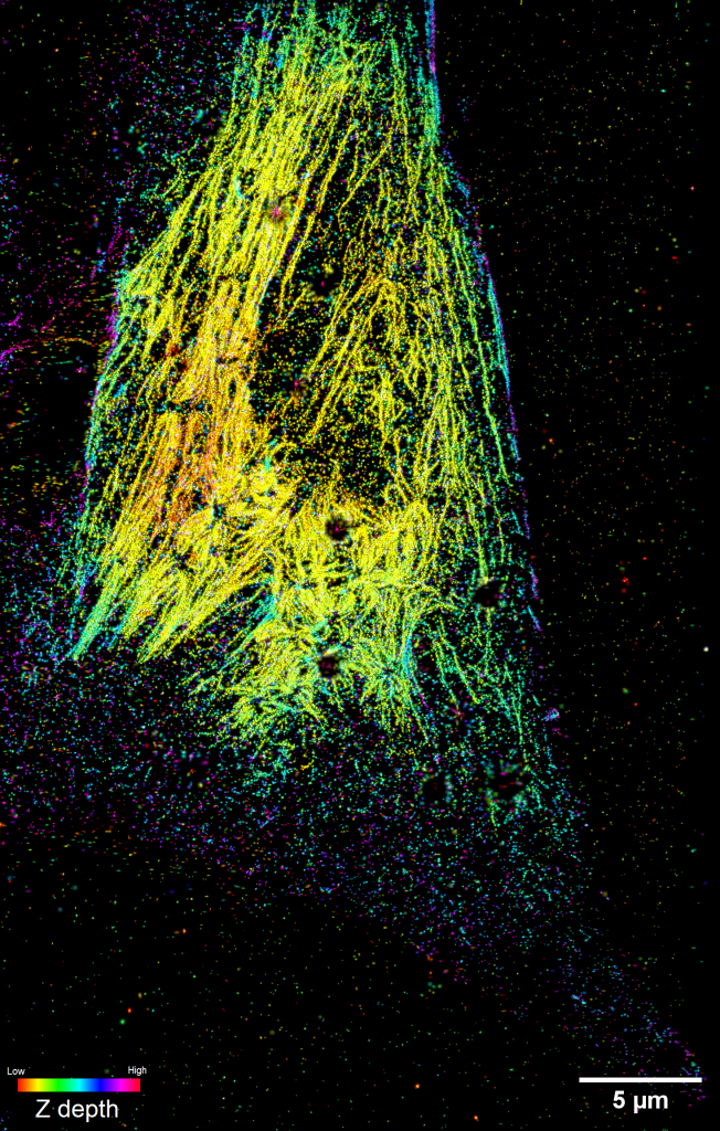
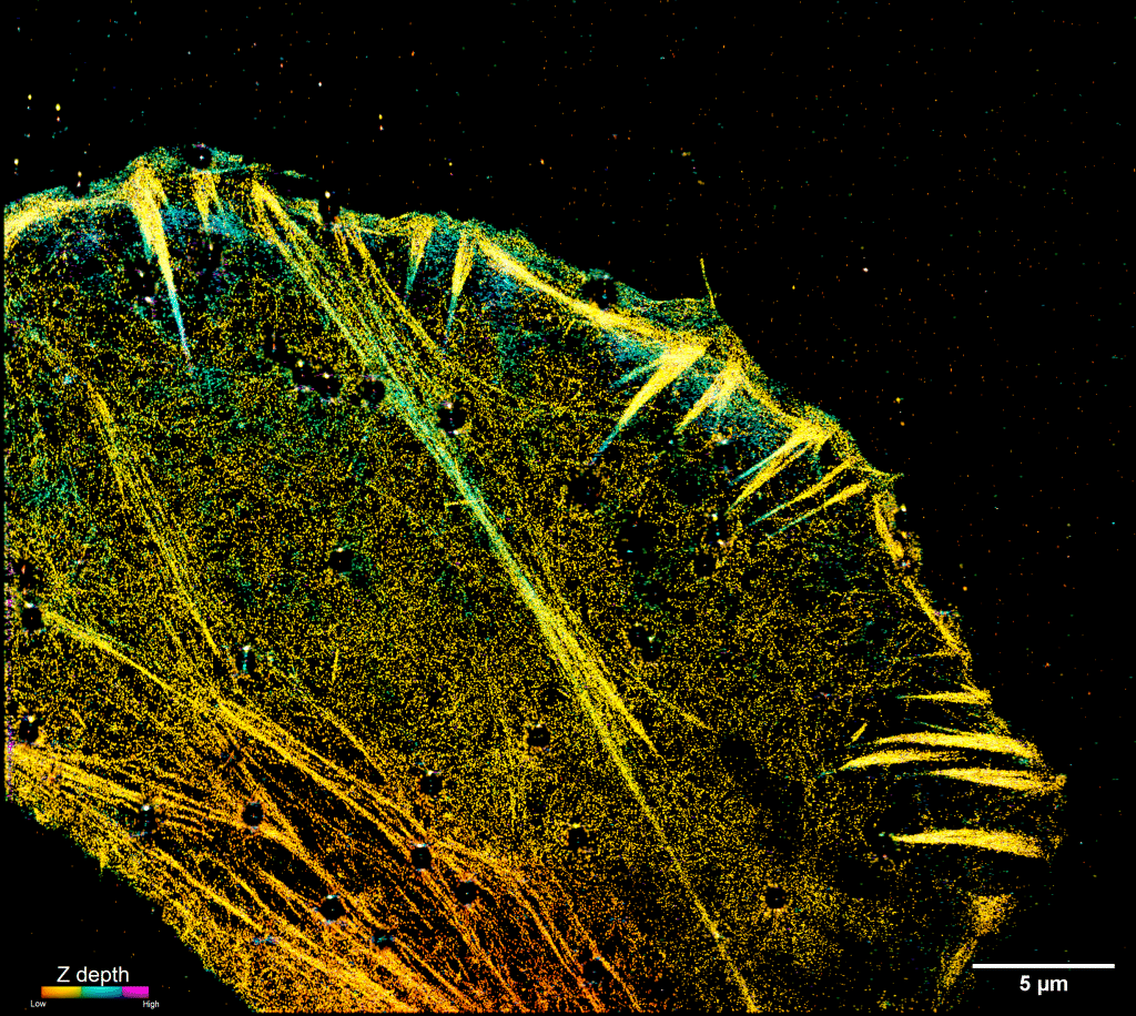
At Janelia, I have further honed my expertise in super-resolution microscopy. During my time as an Advanced Microscopy Fellow, I managed the iPALM, a cutting-edge super-resolution microscope developed in-house at Janelia that combines single molecule localization with interferometry. This enables us to visualize the arrangement of molecules in intricate cellular structures, such as focal adhesions, with exceptional XYZ resolution of 10-20 nanometres. I have assisted other researchers with imaging on the iPALM for several projects that have explored the organization of molecules in various structures, including focal adhesions, muscle fibres, and the cytoskeleton.
Q: Can you tell us about your current roles and responsibilities at Janelia?
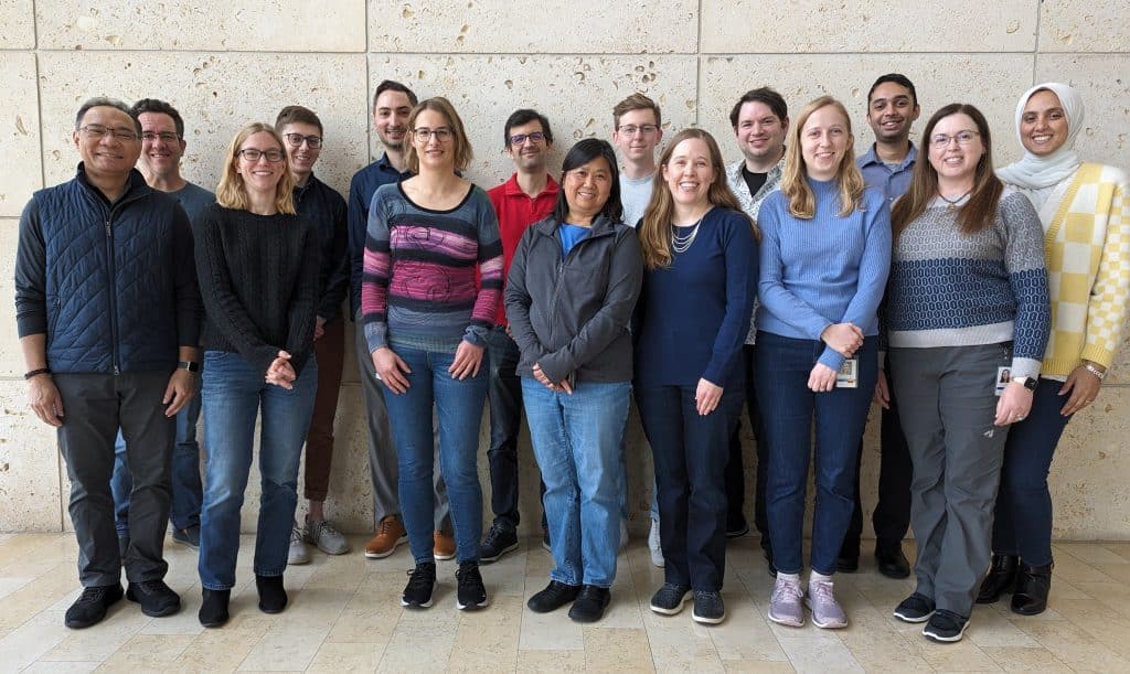
A: I began my journey at Janelia as an Advanced Microscopy Fellow at the AIC, under the mentorship of Dr. Teng-Leong Chew. This postdoc-equivalent training program is specifically designed for individuals seeking to build a successful career in core facilities.
After completing my fellowship, I transitioned from the AIC, which serves external researchers, to the internal light microscopy core facility that focuses on supporting the imaging needs of Janelia’s research community. In my current role, I manage a range of commercial microscopy systems, including confocal and light-sheet systems, and provide support to Janelia’s researchers.
Q: How do you organize training programs and workshops, and what strategies do you employ to ignite scientific curiosity among researchers?
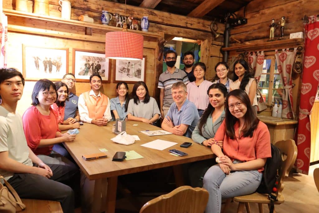
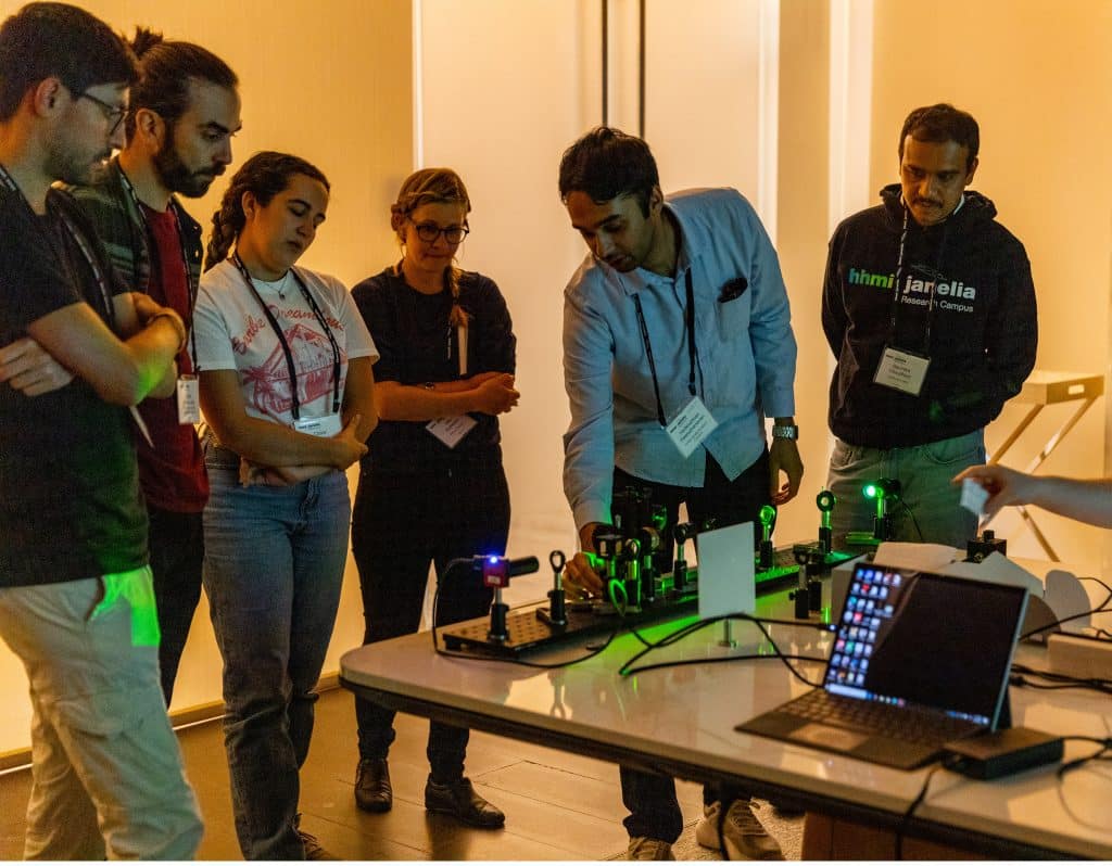
A: We acknowledge our privileged access to cutting-edge microscopy and a wealth of expertise from leading minds available at Janelia. This privilege comes with a deep responsibility to share these resources and uplift researchers in regions where such opportunities are limited. We have previously organized global outreach workshops in Africa, Latin America, and Asia. These workshops introduce participants to the vast and remarkable capabilities of microscopy, emphasizing its quantitative power and unique ability to visualize biological processes in their natural state. In addition to support from Janelia, we have also obtained funding from the Chan Zuckerberg Initiative, the Moore Foundation, and the Gates Foundation, as well as microscopy companies, to bring these initiatives to life.
The workshops typically bring together 24-30 participants from diverse career stages, including PhD students, postdocs, principal investigators, and core facility personnel. We cultivate a collaborative learning environment where participants can network, exchange ideas, and gain hands-on experience with various microscopy techniques. Crucially, the focus isn’t on teaching how to operate microscopes, but rather on how to design and execute hypothesis-driven microscopy experiments to quantitatively address complex biological questions. A large portion of the workshop is also dedicated to data analysis, interpretation, and reporting, empowering researchers to use microscopy effectively to address their research questions.
I recently had the opportunity to organize such an outreach program – the Okinawa Microscopy Workshop – to help foster a collaborative Japanese-Southeast Asian imaging community. The response was overwhelming as we received over 450 applications for just 30 slots, underscoring the significant demand for microscopy in biology research even in developing countries. This high level of interest also highlights the need for initiatives that support and connect researchers across regions, particularly in under-resourced nations where access to such specialized knowledge and resources is often limited. By helping to form regional networks for collaboration, we can facilitate knowledge sharing and enhance the capabilities of scientists in these areas (https://www.nature.com/articles/s41592-024-02397-1).
Q: What’s the most effective approach for an international researcher hoping to collaborate with the AIC?
A: At the AIC, the mission is to empower researchers by providing access to advanced pre-commercial imaging technologies. The first step is to schedule a technical consultation via a Zoom call. This meeting allows you to present your project, highlighting the key biological questions you’re trying to address and the challenges being faced, and justify the need for Janelia’s specialized pre-commercial microscopes over existing commercial options.
If your project is justified and technically feasible for our microscopes, we will ask you to submit a formal proposal. This proposal should be a concise document, approximately 1,000 words, detailing your hypothesis, specific research questions, current challenges, experimental methods, and data analysis plan.
Proposals are reviewed periodically, with calls for proposals occurring 1-2 times per year. If your proposal is accepted, we will arrange a two to three-week visit to Janelia. It is crucial that your project is designed to yield informative data within this timeframe. During this visit, we will provide accommodation, access to microscopes, and necessary reagents. Our interdisciplinary team of experts, including microscopists, biologists, and data analysts, will support you throughout your visit. We are also committed to supporting you beyond your visit, offering advice and assistance with data analysis and interpretation.
Q: In your view, how are the fields of cell biology and microscopy evolving, particularly in light of advancements in artificial intelligence (AI)?
A: Being at Janelia offers a unique vantage point to witness the rapid evolution of microscopy and its application to answer complex biological questions. This is an exciting time for scientists, with a plethora of new techniques and possibilities continuously emerging. We recently published a paper (https://www.nature.com/articles/s42003-023-05468-9) where we envision the future of imaging as one where we can visualize and study everything, everywhere, all at once, with the required microscopy tools being readily accessible to researchers around the world.
AI holds great promise and is poised to revolutionize microscopy research. It can aid in experimental design, automate data collection, processing, and analysis, and extract deeper insights from complex datasets, potentially predicting future testing outcomes. However, it’s important not to get caught up in the hype and AI implementation must be approached with great caution. High-quality training data is essential to ensure accurate, reliable, and reproducible results. When used judiciously and with a strong scientific foundation, AI has the potential to accelerate scientific discovery. HHMI is also investing in AI research to harness its transformative potential effectively.
Q: What advice would you offer to PhD students and young postdocs who are passionate about pursuing a career in cell biology and microscopy?
A: Don’t let a lack of extensive prior experience in microscopy deter you from pursuing your passion. The field is rich with opportunities for learning and growth, and skills and knowledge can be picked up along the way.
Knowing what you don’t want to do is as important as knowing what you do want to do. It’s perfectly acceptable to pivot and explore new directions as your interests evolve. Remember, a PhD provides you with not just specialized technical skills; it also equips you with transferable skills like critical thinking, problem-solving, and effective communication. These abilities are highly valued across different sectors, including academia, industry, and beyond. It’s important to stay open-minded and be flexible with your career trajectory. Embrace the learning process, remain curious, and use your versatile skill set to explore diverse career opportunities.
Q: How do you perceive the current state and future of cell biology and microscopy in India?
A: India has many talented and dedicated researchers. However, despite this strong human capital, the overall scientific progress in the country is often constrained by several systemic challenges. These include limited funding, inadequate infrastructure, restricted access to cutting-edge technologies, and poor collaboration opportunities. This situation has contributed to brain drain, with many researchers, including myself, seeking opportunities abroad where resources and facilities are more readily available.
To realize the full potential of its researchers, India needs to create an environment that retains and nurtures homegrown talent. Building a strong and interconnected local community is essential. Initiatives like ‘India Bioimaging,’ which aim to foster collaboration and knowledge sharing among researchers and core facilities across the country, can play a significant role in this effort. Government and private organizations also must play a vital role in providing the necessary funding, infrastructure, and support for scientific advancement.
It’s understandable that, given India’s large population, it is impossible to fund every lab at the level seen in wealthier nations. The strain on resources is considerable, and it’s simply not feasible to equip every institution with cutting-edge technologies. A more practical and strategic approach (https://onlinelibrary.wiley.com/doi/full/10.1111/jmi.13277) could be the establishment of shared core facilities. Centralized, advanced microscopy core facilities – similar to Janelia’s AIC – could serve as national hubs for high-end imaging technologies and expertise, maximizing the impact of available funding and providing researchers nationwide with easy access to cutting-edge tools.
Q: Have you encountered challenges as an Asian researcher?
A: Personally, I’ve been fortunate to work in supportive environments. However, systemic barriers like unequal access to opportunities and funding persist for many Asian scientists. Cultural biases and stereotypes can also affect perceptions and opportunities, often requiring Asian researchers to work harder to demonstrate expertise and gain recognition. Navigating different academic and professional cultures, especially in international settings, can be challenging, from adapting to diverse working styles to overcoming language barriers.
Moreover, the expectation to balance personal responsibilities with professional achievements can be intense, particularly in cultures where family obligations are highly prioritized. This adds another layer of complexity for Asian researchers, as societal expectations might clash with individual career goals. This creates a delicate balancing act between fulfilling career goals and meeting family obligations, making it even more important to foster supportive work environments that acknowledge these unique challenges. Tackling these challenges often requires perseverance and adaptability, and building a network of mentors and collaborators who understand these unique pressures can be invaluable.
Q: How can the field become more inclusive and supportive of the Asian cell biologist community?
A: To create a more inclusive and supportive environment for Asian scientists, several key steps are essential. Increasing representation within scientific organizations and conferences will ensure that Asian researchers have role models and advocates who understand their specific challenges. Targeted mentorship programs and networking opportunities are vital for building meaningful connections and career support. Institutions should also implement cultural sensitivity training to combat biases and cultivate a more inclusive atmosphere. Supportive work environments that respect diverse cultural practices and family responsibilities will help researchers balance their personal and professional lives. Additionally, recognizing the achievements of Asian researchers through awards and media coverage will enhance their visibility and impact. Finally, fostering strong local research communities will help scientists grow and connect more effectively, while international collaborations will expand their access to expertise and resources.
You can find all the posts from my interview series here.


 (3 votes, average: 1.00 out of 1)
Thanks for the shoutout! This series is a great initiative to highlight Asian scientists.
Very Insightful and motivating especially to us young African scientist