New insights into corneal development from vEM
Posted by Mario Elias Ortega Sandoval, on 17 June 2025
by Rob Young*, Kiranjit Bains*, Phil Lewis* and Andrew Quantock*
*Biophysics Research Group, School of Optometry & Vision Sciences, Cardiff University, UK
Challenge
The unique transparency of corneal stromal tissue, essential for vision, is dependent upon the remarkable order of component collagen fibrils, which exhibit highly uniform diameter, spacing and orientation. However, processes involved in its formation during embryonic development remain unclear. A better understanding of stromal damage and repair in disease and healing is also a major challenge. Much of our knowledge stems from observations made using conventional 2D imaging methods, but the advent of high-resolution volume EM has opened up the possibility of extending our understanding into 3 dimensions to reveal novel structures with unknown function and to validate the avian eye as a model experimental system.
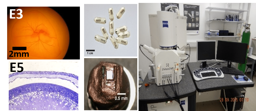
Technique: serial block-face (SBF) SEM
Corneal tissue from developing avian eyes at different embryonic (E) stages is fixed in aldehyde solution, contrasted with heavy metals, dehydrated and embedded in epoxy resin. Blocks on specimen pins can then be trimmed, polished and sections stained with toluidine blue for orientation and selection of regions of interest. Datasets of ≤ 1000 serial images at 100 nm intervals and 5-10 nm/px resolution are acquired in a scanning EM equipped with an ultramicrotome. Datasets are aligned, converted to tif format and segmented using specialist analysis software to generate 3D reconstructions of cell / matrix interactions in large tissue volumes.
Research
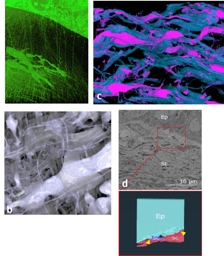
Our studies show early keratocyte migration forms the corneal stroma, as surface epithelium sheds microvesicles (evs) and extends matrix cords into the underlying primary matrix (a); cells express elongate keratopodial processes involved in alignment of orthogonal lamellae of secreted collagen fibrils (b,c); direct stromal- epithelial cell interactions (d) across the basement membrane at the limbus (arrowheads) suggest this is the site of the limbal stem cell niche.
Ep – Epithelium, St – Stroma, SC- Stromal cell.
Impact
• Stroma 3D data informs synthesis of corneal replacements for therapy
• Studies of cell-guided matrix deposition and evs role in development
• Avian eye as a model for studies of the limbal stem cell niche
References
(1) Bains et al. Cells 2023 doi.org/10.3390/cells12192334
(2) Young et al. 2020 Arch Clin Exp Ophthalmol doi:10.46439/ophthalmology.2.014
(3) Young et al. 2019 Exp Eye Res. doi: 10.1016/j.exer.2019.107772.
(4) Young et al. 2014 PNAS doi: 10.1073/pnas.1313561110
Poster
 (No Ratings Yet)
Get involved
Create an account or log in to post your story on FocalPlane.
More posts like this

Filter by
- NewsApply
- DiscussionsApply
- How toApply
- ToolsApply
- Case studiesApply
- InterviewsApply
- JobsApply
- EducationApply
- Blog seriesApply
- Volume EMApply
- Latin American Micro..scopistsApply
- Bio-image Analysis w..ith NapariApply
- Imaging with…Apply
- Towards Global Acces..sApply
- Latin America Bioima..gingApply
- From Zero to Qupath ..HeroApply
- Asian Microscopists ..and Cell BiologistsApply
- AIC at HHMI JaneliaApply
- Deep Learning for Bi..o-image analysisApply
- GloBIAS – updates fr..om the communityApply
- Highlights from Euro..-BioImagingApply
- LSFM seriesApply
- DIY MicroscopyApply
- View all