Elisabeth Kugler is the director of Zeeks - Art for Geeks Ltd. A company transforming the way you think about data. We are passionate about microscopy data, how to analyse, visualize, and communicate them. Previously, she was a PostDoc at UCL, developing a quantitative model of retina neurovascular unit formation. She conducted her PhD at the University of Sheffield, developing image segmentation, registration, and quantification pipelines for the zebrafish cerebral vasculature. During her PhD she discovered and characterized a previously undescribed cell membrane behaviour, named kugeln. Elisabeth is passionate about all things science and art. In her free time, she collects what someone may call too many books, fossils, and rocks.
Scientific field: Developmental biology, Computational biology
Microscopy background: Image Analysis
Posted by Elisabeth Kugler, on 20 May 2025
Posted by Elisabeth Kugler, on 8 September 2023
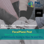
Reflections and Revelations: Highlights from #PoLBIAS23 - A Symposium on Bioimage Analysis. This blog showcases a summary, as well as a reflection on open questions and community efforts,.Posted by Elisabeth Kugler, on 26 July 2023
Posted by Elisabeth Kugler, on 12 June 2023
Posted by Elisabeth Kugler, on 17 May 2022