
Advertisement

Displaying posts in the category: Case studies

Imaging spotlight: the axonal membrane-associated periodic skeleton

Posted by , on 5 March 2026

In this paper highlight, Nick Boyer takes us through the key results and imaging techniques that they used in their research dissecting the axonal membrane-associated periodic skeleton with Rohan Sharma, Christophe Leterrier, Subhojit Roy and colleagues.

Imaging spotlight - How to divide without a ring: a mechanical ratchet drives unilateral cytokinesis

Posted by , on 10 February 2026

In this paper highlight, Jan Brugués and Alison Kickuth discuss the microscopy and microrheology techniques that they used in their research dissecting the mechanics of cytokinesis.

Seeing Chromatin Scaling with Cell Size, from Interphase to Mitosis

Posted by , on 26 January 2026

Melike Lakadamyali, Jérôme Solon and Manuel MendozaAs cells divide during development, their size evolves dramatically while DNA content remains constant. This raises a fundamental question: does chromosome compaction adjust to these size changes so that genomes are still segregated faithfully? This question has been with us for a long time. More than a decade ago,

Imaging spotlight: visualising synaptic vesicle fusion by cryo-ET

Posted by , on 21 January 2026

In this paper highlight from Jana Kroll and colleagues, we hear about their research combing optogenetic stimulation with plunge freezing and cryo-electron tomography to study synaptic vesicle fusion.

Imaging spotlight: U-ExM atlas of microbial eukaryotes

Posted by , on 20 January 2026

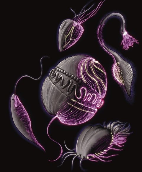
In this paper highlight from Hiral Shah and colleagues, we learn about their ultrastructure expansion microscopy (U-ExM) atlas of over 200 cultured planktonic eukaryotes. This post includes technical advice for ExM users and highlights the open-access resource that they have generated for cytoskeleton or marine microorganism aficionados (and microscopy sciart lovers!)

Imaging spotlight: Initiation of chromosome congression deconstructed by multi-scale live lattice light-sheet and super-resolution microscopy

Posted by , on 16 January 2026

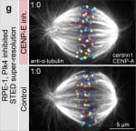
In this highlight, Kruno Vukušić and Iva M. Tolić describe how they use multi-scale live lattice light-sheet and super-resolution microscopy to decipher the earliest steps of chromosome congression.

Imaging spotlight: Revealing mouse brain ultrastructure using non-destructive X-ray tomography

Posted by , on 13 January 2026

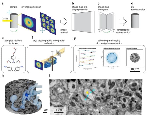
In this paper highlight, Carles Bosch, Ana Diaz, Adrian A. Wanner and Andreas T. Schaefer (on behalf of all authors) describe how they use nondestructive X-ray tomography to map mouse brain tissue ultrastructure . 

Damage done, tubules on - Studying membrane damaged-induced lysosomal tubulation 

Posted by , on 11 December 2025

In this post, I'll told you the "ins and outs" of our last publication in JCB about lysosomal tubulation in response to damage.

Epithelial Mechanics Fan Club

Posted by , on 4 December 2025

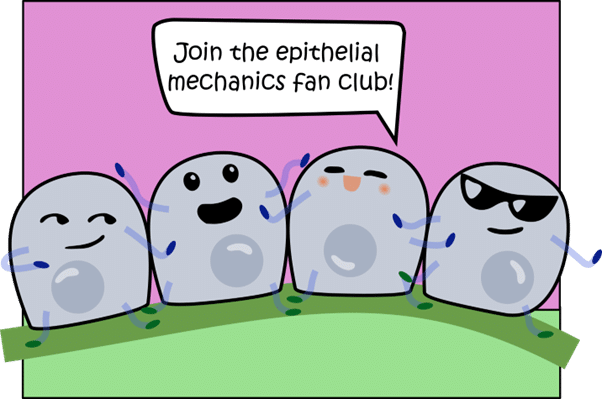
In this post, Nimesh Chahare and Julia Eckert tell us about the 'Epithelial Mechanics Fan Club'. You can follow the club on Bluesky, X, and now on their new website. Reach out to Nimesh and Julia if you would like to contribute.

An Introduction to the Croatian Microscopy Society

Posted by , on 19 November 2025

The origins of the Croatian Microscopy Society (CMS) can be traced back to 1983, when the Section for Electron Microscopy was established within the Croatian Society of Natural History at the initiative of Prof. Zvonimir Devidé, the first head of the Section. What began as a small group of enthusiasts meeting in Zagreb to exchange