Advertisement

Displaying posts in the category: News

The FocalPlane Network: joining and using the database

Posted by , on 20 September 2023

Our instructional videos take you step-by-step through the process of joining and using the FocalPlane Network. You can contact us at focalplane@biologists.com with any feedback on the Network. Joining the FocalPlane Network Using the FocalPlane Network

The FocalPlane Network: connecting the microscopy community

Posted by , on 20 September 2023

The FocalPlane Network is an international directory of researchers with expertise in microscopy. The goal of the Network is to facilitate promotion and networking, as well as assisting those seeking conference speakers, committee members, reviewers or collaborators. Membership is open to researchers in the microscopy community from all career stages, geographical locations or areas of

Featured image with Martín Estermann

Posted by , on 15 September 2023

Our featured image, acquired by Martín Estermann, is a 3D reconstruction of C. elegans transgenic embryos. Embryos were imaged live using a Zeiss Celldiscoverer 7 microscope. The 3D reconstruction was performed using Fiji. Histones in are shown in green and the cell membrane in magenta. We caught up with Martín to find out about his

And the winner is...

Posted by , on 1 September 2023

We are delighted to announce that the winner of the 2023 FocalPlane image competition is Rebecca Simkin. Rebecca’s image, ‘Neuromuscular junctions’, depicts four NMJs in a lumbrical muscle, located in the hind paw of a wildtype mouse. A wholemount immunostaining was performed using 2H3 and SV2 antibodies to visualise lower motor neurons in green (specifically

Unveiling the Marvels of the First-Ever Gordon Research Conference on Volume Electron Microscopy - Capturing Subcellular Architecture from Organelles to Organisms -By Ana Correia and James Thompson

Posted by , on 24 August 2023

We delve into our captivating journey at the first Gordon Research Conference (GRC) on Volume Electron Microscopy (vEM) as early career scientists. Set against the backdrop of sunny Ventura, California, this conference brought together experts in the vEM field from all corners of the globe, creating an intellectual haven for learning, networking, and forging collaborations.

Featured image with Beste Senem Değirmenci

Posted by , on 18 August 2023

Our featured image, ‘Bubbles of The Cell’, shows enlarged endosomal vesicles in HeLa S3 cells upon inhibition of PI(3,5)P2 activity. PI(3,5)P2 is a crucial phosphoinositide involved in the regulation of endosomal trafficking. Depletion of PI(3,5)P2 disrupts the transition from early to late endosomes, leading to the enlargement of endosomal vesicles. Imaged with Leica DMI8 SP8

Featured image with Outi Paloheimo

Posted by , on 4 August 2023

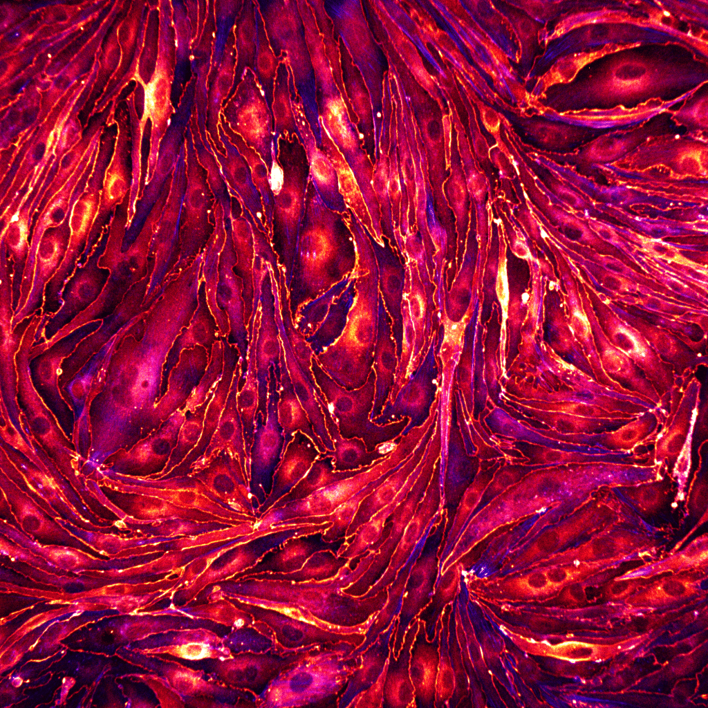
Our featured image is laser scanning confocal microscope image showing cell division of Madin-Darby canine kidney cells with nuclei in grey and filamentous actin in orange. The image was acquired with a Zeiss LSM 780 microscope with a 63x/1.40 oil immersion objective, 405 nm diode laser and 561 nm diode-pumped solid-state laser. Deconvolution was performed

The FocalPlane correspondents: Marta Forcella

Posted by , on 26 July 2023

At the beginning of the year, we announced that we will be working with three FocalPlane correspondents, who will be helping us to develop and write content for FocalPlane. We caught up with each of them to chat about their research backgrounds and the topics that they’re excited to write about. Here, we talk to

Featured image with David McGrath

Posted by , on 21 July 2023

Our featured image is of mature mouse brown adipocytes (WT-1 cell line) under normal conditions from David McGrath. Channel 1 (green LUT) shows lipid droplets stained with BODIPY 493/503 and channel 2 (fire LUT) shows mitochondria stained with TMRE. The image was obtained using a Visitron SD-TIRF confocal microscope (60x Apo TIRF (corr.) Oil/ NA: 1.49) at the UKE Microscopy Imaging Facility

New on the Node — Unveiling the Unseen: Organizing a Science Art Exhibition

Posted by , on 12 July 2023

What happens when one combines their love for art, microscopy and developmental biology? In a blog post on the Node, Elisabeth Kulger talks about her experience of planning a science art exhibition showcasing the intricate world of developmental biology through the lens of microscopy. Head over to the Node to read more about the challenges,