Advertisement

Displaying posts with the tag: is_archive

Please sign and share: open letter to Nikon on the 2026 Small World in Motion winner

Posted by , on 8 October 2026

Dear microscopy friends and colleagues, You may have seen that the 2026 Nikon Small World in Motion competition was won by a video captioned as showing ‘abnormal beating of airway cilia from a child with primary ciliary dyskinesia‘. It has since been shared by major news outlets worldwide. The submitter has said that the regions

FocalPlane features... cell migration recordings with Ankita Jha, Raphaël Clément and Katrina Velle

Posted by , on 25 September 2026

On 17 September 2026, we held the second webinar in our FocalPlane features… series on emergent models and quantitative analysis of cell migration, organised by Pablo J. Sáez and Valeria Venturini. In this webinar, we heard from Ankita Jha on ‘Decoding bleb driven signaling in cancer disease progression’, Raphaël Clément on ‘Navigating without neurons or

JCS snapshots: the cilia and flagella edition

Posted by , on 24 November 2025

Journal of Cell Science recently published a Special Issue on Cilia and Flagella: from Basic Biology to Disease, edited by Pleasantine Mill and Lotte Pedersen. We invited the authors of the research published in this issue to produce JCS snapshots, short videos highlighting the key findings from their papers. Below, you can find the snapshots

JCS Special Issue - 'Cilia and Flagella: from Basic Biology to Disease'

Posted by , on 10 November 2025

We'll be celebrating the research published in JCS's special issue on cilia and flagella with the UK Cilia and Centrosome Network at their upcoming symposium on Tuesday 25 November.

Featured image with Kaiden Power

Posted by , on 26 September 2025

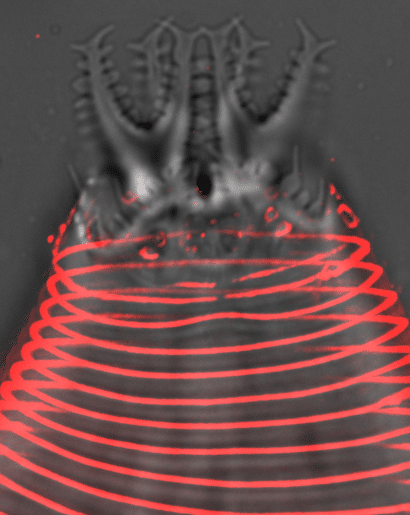
Our featured image, acquired by Kaiden Power, shows the head and probolae of an Acrobeles complexus nematode. The cuticle furrows (annuli) and cilia pores are stained with Concanavalin A (red). The sample was mounted and imaged live on a DeltaVision widefield microscope with deconvolution post-acquisition.

Microscopy preprints – applications in biology

Posted by , on 19 September 2025

Here is a curated selection of preprints published recently. In this post, we share preprints that use microscopy tools to answer questions in biology.

Featured image with the Hughes lab

Posted by , on 20 December 2024

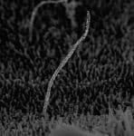
Our featured image, acquired by Sanja Sviben, is an immuno-scanning electron micrograph of a primary cilium, taken from an intact human pancreatic islet. The cilium is a microtubule-based structure and is seen here labeled with colloidal gold for acetylated alpha tubulin.