Get involved
Create an account or log in to post your story on FocalPlane.
Microscopy-related articles from our journals
- Toggle-Untoggle – a cell segmentation tool with an interactive user verification interface J Cell Sci 2025 138: jcs264154
- The ratio of Wnt signaling activity to Sox2 transcription factor levels predicts neuromesodermal fate potential Development 2025 152: dev204661
- The short isoform of Tango1 is dispensable for zebrafish survival but is required for skeletal patterning and integrity Biology Open 2025 14: bio062117
Microscopy-related preprint highlights

- When geometry isn’t enough, actin helps plant cells decide where to divide, ensuring robust tissue patterning
- Planar cell polarity protein, Vangl2, goes out of its way to shape the heart through a planar polarity-independent mechanism.
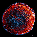
- HAK-actin, U-ExM-compatible probe to image the actin cytoskeleton


