Featured image with Sujan Ghimire
Posted by FocalPlane, on 26 April 2024
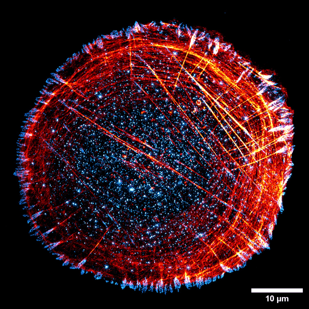
Our featured image, acquired by Sujan Ghimire, shows a U2OS cell stained for paxillin (Cyan Hot) and actin (Red Hot) following an immunofluorescence staining protocol. The image was captured using structured illumination microscopy (SIM) with Deltavision OMX (63x Oil, NA: 1.42) at Cell Imaging and Cytometry facility, Turku Bioscience. After SIM reconstruction, the maximum intensity projected image was processed in Fiji to generate the final image.
Find out more about Sujan’s research below:
Research career so far: I am a third-year doctoral researcher at the Cell Migration Lab (https://cellmig.org) at Åbo Akademi University in Turku, Finland, under the supervision of Associate Professor Guillaume Jacquemet. During my second year in the Master’s Degree Programme in Biomedical Imaging (BIMA) at Åbo Akademi University, I joined the lab as a summer intern in May 2020 to work with Dr Gautier Follain. Following the internship, I continued with my master’s thesis and doctoral research project in the lab.
Current research: Currently, I am investigating pancreatic cancer metastasis, where tumor cells from primary tumors migrate towards the blood and lymphatic vessels to circulate throughout the body as circulating tumor cells. These cells then undergo arrest in circulation before eventually extravasating from the bloodstream to metastasize in distant organs. I am focused on elucidating the mechanisms underlying the arrest of circulating tumor cells at distant organ sites and their subsequent extravasation from blood vessels using a variety of imaging modalities, ranging from brightfield to super-resolution microscopy, on both live and fixed samples.
Favourite imaging technique/microscope: I am passionate about microscopy and like all microscopes, from brightfield to super-resolution and electron microscopes. Each microscope is designed to suit specific samples and varies in resolution, yet all serve the purpose of visualising the invisible. Throughout my microscopy career, I have found particular joy in live-cell imaging in super-resolution using LSM-880 Airyscan.
What are you most excited about in microscopy? The field of microscopy is evolving rapidly, with several different super-resolution microscopy techniques being developed. In addition to optics and image reconstruction-based super-resolution microscopy, expansion microscopy is also gaining popularity. I am particularly excited about the advancements in live-cell imaging in super-resolution, and image analysis tools. With the increasing popularity of AI in recent years, the microscopy and bioimage analysis fields will surely see steep upgrades.
 (No Ratings Yet)