Career insight: bioimage analysis
Posted by Pradeep Rajasekhar, on 3 July 2020
I am a postdoctoral fellow based at the Monash Institute of Pharmaceutical Sciences (MIPS), Melbourne, Australia. I am originally from Kerala, India and travelled to Australia for my postgraduate studies almost 13 years ago. I came to Melbourne because of all the high quality and affordable educational opportunities, and fell in love with it for its vibrant culture, coffee, and its great culinary scene. It is where I met my amazing partner, and it is the place that I now call home.
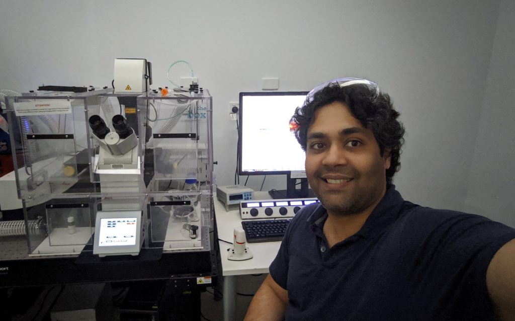
Currently I work in the Integrated Neurogenic Mechanisms (INM) Laboratory at MIPS, and we focus on characterising drug targets in pain and inflammation. What I really like about the lab is that we examine biological processes from cells in a dish, to cellular interactions in tissues and all the way to circuits in whole-animal models. My research focuses on the network of nerves within the gut, the enteric nervous system (ENS), which controls how the gut functions and is often referred to as the ‘second brain’ of our body. I work on an industry-funded project where we utilise microscopy and bioimage analysis (BIA) to characterise the cellular environment within the gut, focussing on neuro-immune interactions. The project is a perfect combination of the academic and industry worlds. It focuses on basic biology, whilst at the same time involves interacting with clinicians and industry leaders in solving real-world medical problems.
My passion for the biological sciences and interdisciplinary research goes back to my original training in Biochemical Engineering. It was during my degree that I had my first ‘wow’ moment in Microbiology, where we immersed a blade of grass in nutrient broth, left it overnight and looked at it under a light microscope using the hanging-drop preparation. It was amazing and exciting to see a entire new world inside that small liquid drop. My interest in microscopy continued to grow during my PhD where I spent countless hours taking images of cells (fixed and live) in cell culture dishes and whole tissue samples. As much as I enjoy looking at these beautiful images, being a scientist, I believe that beauty is also in extracting meaningful information from these images to present a story of what is happening at a microscopic level.
My PhD training gave me invaluable training in experimental design and exposure to the whole bioimaging research workflow, i.e. from wetlab experiments and image acquisition, to image processing and data analysis. As the research questions became more complex, I found myself spending increasing amounts of time on image and data analysis. We know that it takes so much time, effort and money to generate research data. Why not put the same effort into analysis and get a deeper insight into the biology? I realised that bringing together the fields of biology, microscopy and image analysis is what I enjoy doing and is where I can add value.
Although I have been spending a significant amount of time analysing biological images for the past 2 to 3 years, I never really thought of myself as a ‘Bioimage Analyst’ (BIAlyst). In fact, I did not even know that the term existed until 2 years ago. A BIAlyst specialises in developing image analysis (IA) workflows by assembling, adapting and automating multiple computational tools, to quantify biological phenomena in an unbiased and scientific manner. They form a bridge between life scientists and software developers, aiding in the development of tools that reveal novel biological insights [1].
My understanding of the biology, and my engineering background together helps me:
- Identify and provide input about the appropriate choice of experimental techniques
- Understand the types and modalities of information that can be extracted
- Explore and identify the right tools across multidisciplinary fields
- Apply the right computational tools to quantify and interpret the data
- Identify and collaborate with the right people from other disciplines so that skills and knowledge can be transferred and applied to solve biological problems
This is summed up perfectly in an article that I read about bridging quantitative disciplines and cell biology: “Having insights into where to look, what to look and how to look” will help maximise research output and impact. As a BIAlyst, I can contribute to the efficient design of experimental workflows in their entirety. For example, a major part of my work involves live imaging of gut tissue wholemounts. Depending on the aim of the experiment, I can provide input on the type of tissue preparation, choice of treatment conditions, and the modality and acquisition settings of the microscope in order to maximise the amount of information extracted.
I was fortunate enough to attend the Network of European Bioimage Analysts (NEUBIAS) 2020 meeting. NEUBIAS has been leading the way in generating awareness and recognition of BIA as a career option. They are fulfilling the need for more training opportunities through their training schools and the NEUBIAS Academy@Home online workshops. NEUBIAS 2020 was an educational and humbling experience where I interacted with leading experts, learnt about cutting edge tools and software, and became a part of a community of like-minded professionals. I am currently applying the techniques I learnt at this conference in my research projects and hope to use my new network to develop my skill-sets and initiate new collaborations.
The BIA community is well-developed and recognised in Europe with initiatives like NEUBIAS, and in the USA where the Center for Open Bioimage Analysis (COBA) was established in 2020. We have a strong microscopy community in Australia, but BIA as a field and career option is still not very well recognised. Almost 70% of high-impact bioscience publications use advanced microscopy techniques, and Australian universities invest millions of dollars in acquiring high-end microscopes. This is not supported well enough with the capacity to analyse the large amounts of data being produced. For example, at Monash University we have only four people who officially do BIA (amongst other responsibilities) for a user-base that is easily in the 1000s.
Although there are plenty of free and paid software packages available, they either have numerous components or tools that require a basic understanding of image processing algorithms, or they are too specific for a niche biological problem. This is where a BIAlyst can add value by assembling and customising IA workflows to answer specific research questions. This ensures better return on an investment of government research funding by maximising the research output and productivity. For this to happen in Australia, it is imperative that we develop a strong community of BIAlysts where we can share our knowledge and expertise, as well as increase our representation in local and international meetings. I hope that in the future we can have national networks and research centres for open BIA in Australia, and I look forward to being an active member of this community.


Pradeep Rajasekhar
Postdoctoral Fellow
Monash Institute of Pharmaceutical Sciences (MIPS)
Melbourne, Australia


 (No Ratings Yet)