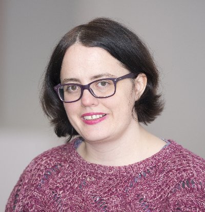
‘Under the scope’: an interview with Ann Wheeler
Posted by FocalPlane, on 8 September 2020

Ann Wheeler received her PhD from University College London as part of a molecular cell biology 4-year rotation programme where she got to use confocal imaging quite extensively. She then moved to Columbia University to do some total internal reflection fluorescence (TIRF) microscopy. She was actually one of the first people to use TIRF as there was no biological system for doing TIRF imaging in the UK at the time. Ann was really interested in applying advance imaging techniques to biological questions so she then joined Clare Waterman’s lab where she did speckle fluorescence microscopy, TIRF and spinning disc imaging. She then came back to the UK to work on a high content screen at Imperial College London. With a paper in the making and a project left unfinished due to funding issues, Ann was then offered a position at Queen Mary University London as a core facility manager where she stayed for 5 years. Since then, she moved to Scotland and joined the Institute of Genetics and Molecular Medicine (IGMM) to run the core imaging facility. She’s been there for the last 5 years, managing and providing imaging services for the institute and external users.
You started in molecular cell biology but then moved on to microscopy. How challenging was it to switch?
I didn’t find it difficult at all. I still consider myself a biologist, my training is in biology. I was working on cell motility and I have an ongoing interest in cell motility in metastases. One of the reasons I accepted this job here is because I am able to be involved in projects involving molecular genetic disease and cancer research. What I do and have always done is visualise the process whereby cells migrate, move, reorganise their structures in response to various disease situations that could be genetic or drug challenge or a virus. Imaging tools allows researchers to better understand the spatial and temporal mechanisms behind these processes. I would say ‘the switch’ was easier as I have a bit of a physics background and all the labs that I worked in use microscopy quite extensively as a tool. I worked with a lot of postdocs that were very experienced and had some good relationships with industrial partners. I had also received excellent training by my PhD supervisor Anne Ridley. Having worked as a postdoc with Clare Waterman I was able to meet excellent world-leading imaging people like Jennifer Lippincott-Schwartz, Ron Vale and others. I am very grateful for the people that changed me because I think they went out of their way and didn’t need to do that and that was just lovely for me.
You mentioned that you have a bit of physics background: do you think that’s essential to have that background to make the change to microscopy?
It is important to understand the physics and the optics. I studied physics all the way through to the first year of university and that was quite important. In certain respects my undergraduate degree was you could say a little bit old fashioned because they still insisted on teaching people physics and chemistry in the first year and a lot of courses these days they really don’t do this anymore. However for the job I have now, really understanding the optics is quite important. It is possible to learn it after but if you have not studied it since school it’s a lot of ground to cover. Some of the advanced techniques I use like structured illumination microscopy you have to understand wave-particle duality, otherwise the way maths and the techniques work really don’t make sense at all. If you are just going to turn on the instrument and press a button and get an image then you probably wouldn’t need all of that understanding but if you are really going to get into the technique and learn how to truly apply it well to scientific question you do need to understand some physics helps. These days there are a lot of opportunities to learn physics using online courses such as Coursera so it is much easier to get the knowledge that you need than it was when I was studying in the 90s or early 2000s. Now it’s just a case of putting yourself in a place where you can access it.
What’s your day-to-day work at the facility like?
*laughs* Well it varies. No two days are the same. So what I do is I will work with PIs, postdocs and students to really apply the imaging knowledge that I have, maybe more the physical sciences, to the projects they are doing. I work with one student, for example, to set up a new methodology that has never been done before to visualise macrophages moving out of the bone and cancer cells moving with the macrophages. That’s quite interesting because you’ve got to develop the whole workflow from sample preparation, the imaging itself and the quantification of the imaging. While doing that I am also working on a grant application at the moment because we are working to see whether we can get the funding or not to make a big investment for the next generation of our super-resolution microscopy equipment. So I’ll be spending some of the day working with the various academics and support teams to make sure that everything’s in place for that grant application. Our website needs work – as ever so some time will be for that. Meeting one-to-one with my team is very important. We’ve invested quite a lot of money in conditional imaging so I will be chatting with the guy on my team who is an expert on that. He’s setting up the background and the programming background for the conditional imaging and my image analyst is setting up an online resource to help people to access what we’ve done and what we’ve provided. We want to put what we’ve done in terms of our analyses resources out there to the community but we need to organise it before, so I will have to check on that… So quite busy! *laughs*
So what was the most challenging thing you had to face when giving advice?
I think in some respects it’s managing expectations because imaging is one of the sweetest tools that a biologist will need to use. [But] you can’t use imaging for everything and imaging is not going to answer all your molecular biology questions. You do need to use other techniques. So even with super-resolution, it’s not cryo-EM, the throughput is considerably slower and so that can be challenging managing people’s expectation of what they are going to get. There’s also a cost implication as well; we charge for the use of our equipment. There are all sorts of different ways to answer a biological question but sometimes is the case of finding methodologies of best value; maybe we won’t get the best resolution but actually get the right number of repeats for the analysis people are doing then that would actually give them something better than if they’d done the STORM experiment which they would only be able to do once. Sometimes it’s frustrating because we have a lot of instruments and some of them are new to the market but that means they are not always the most stable. For other applications such as bone cancer metastasis imaging, that’s never been done before and the instrument that we are using hasn’t actually been designed to do that job so we are tweaking it a bit to make it work. So obviously that’s frustrating for the researcher because the microscope might stop after doing a lot of great work and get halfway through and the whole system crash because the dataset is too big. That can be a bit frustrating for everybody because you were expecting something really great and then you have to do a bit more work in order to make it to what it’s required for publication.
If you had to give one piece of advice to other microscopists or experts on communicating with a non-expert what would it be?
If you watch ‘The Apprentice’ they talk about understanding your customer and the market. And I think that’s really important actually. Our team try to attend our internal seminars weekly so we can find out what the PhDs and the postdoc are doing. If there’s somebody who uses the facility fairly often we make sure that we go to their talks so we could often chat with them afterwards to find out how things are going and if we have support their work better. We stay in good relationships with what we call our super-users. Our super-users are postdocs and PhD students that use our facility very often and in a way they can act like a bridge because they are the people in the lab and so if there’s a person who doesn’t use the facility and they are a bit concerned about it we’ve got various super-users throughout the institute and they are pretty well-known because they are usually doing a lot of imaging and talk a lot and are socially active. They are helpful as they can reassure researchers with less experience in imaging that the work is feasible and give them some tips.
For most of our facility users, they want to focus on the science, imaging is a tool that’s in the toolbox and there are other tools as well. So you’ve got to remember people that want to do imaging and everything else it’s important to make it quite smooth as an experience to them.
And what piece of advice what you give to the non-experts then?
To the non-experts I would just say “please come and ask. Please don’t assume that you don’t have the knowledge, you don’t have the skills”. I’ve taught people who’ve never touched a microscope to do super-resolution. They were virologists and sort of standard microscopy wouldn’t help them because viruses are too small to be seen but I’ve actually taken people from zero to publishing really good papers in imaging. And they’ve done all the microscopy themselves. These projects can be a lot of fun because the researchers realized they could get scientific results they’ve never seen before and nobody else had seen before because they were willing to just be brave and take a step. I guess that’s what I would say “be brave and take a step” because there are definitely people out there in the community who really want to help you get the best from your science and will enjoy doing it as well.


 (5 votes, average: 1.00 out of 1)