Meet the speakers: Hard X-ray Imaging of Biological Soft Tissues symposium
Posted by Carles Bosch, on 29 September 2021
The Francis Crick Institute is hosting the 2nd edition of ‘Hard X-ray Imaging of Biological Soft Tissues symposium’ on Thursday 14 October 2021, 12:00-18:35 BST. In a series of posts on FocalPlane, we will introduce the speakers participating in this symposium. We hope you find some or all of these talks interesting and you join us on Thursday 14 October.
Anne Bonnin “Multiscale and ultra-fast biomedical applications at the TOMCAT beamline (SLS)“
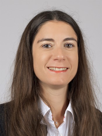
Anne Bonnin is currently Beamline Scientist at the TOMCAT Beamline (SLS, PSI, Switzerland). She received her PhD in Physics from the National Institute of Applied Sciences of Lyon (INSA Lyon, France) in 2009. After four years in the X-ray imaging group at the European Synchrotron Radiation Facility (Grenoble, France), she joined as post-doc in April 2014 the SLS X-ray tomography group and became Beamline Scientist Tenure Track in January 2016. Specialized in X-ray imaging (micro and nano-tomography, single-distance phase-retrieval, diffraction tomography), her research interests focus on the development of new imaging methods to characterize materials and understand their behavior at different length and temporal scales. More particularly, she is in charge of the TOMCAT nanoscope, a full field transmission X-ray microscope (TXM). Since 2016, she leads the Heart Imaging Project at TOMCAT, part of the international collaboration with the UPF, UCL and UHCZ team, aiming at better describing and understanding the cardiac remodelling processes in rat models and human hearts. The various technical advances of this project are now used for other projects in the biomedical field.
Talk description: The recent technical developments carried out in absorption and propagation-based synchrotron X-ray phase-contrast tomographic imaging (PB X-PCI) grant access to high spatial and temporal resolution. This imaging technique is well known to provide 3D details with the great advantage to be non-destructive, without cumbersome sample preparation, and with real-time tracking ability. With the growing interest in studying hierarchical materials, like in the biomedical field, it is crucial to observe samples at different length scales and even by combining different analysis techniques. This talk will present the current multiscale X-ray imaging capability and how this has fostered the (in-vivo) bioimaging program in our group.
Kara Fulton “High-throughput ultrathin serial sectioning method for electron microscopy”
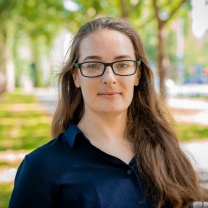
Kara Fulton is a postdoctoral fellow at Harvard Medical School in the department of Neurobiology. She holds a Ph.D. in neuroscience from Brown University and a bachelors degree in mathematics from the University of Michigan. Her Ph.D. research, conducted in the lab of Dr. Kevin Briggman at the Caesar Research Center, included developing methods for correlative electron microscopy and high-throughput serial section collection, and she investigated the functional and structural wiring specificity of interneurons in the mouse olfactory bulb. Her research interests include sensory neuroscience, correlative microscopy, and mathematical modeling of neural circuit computations.
Talk description: Volume imaging of large brain areas with the resolution necessary to reconstruct anatomical connectivity is currently achieved with electron microscopy. Sections are either acquired serially prior to imaging or by iteratively imaging and sectioning within the vacuum chamber of the microscope. To address the technical limitations of serial-sectioning, we have developed an alternative high-throughput non-destructive method for collecting ultrathin sections from large volumes. Compared to the current state-of-the-art, our method provides a robust pipeline for sample preparation, image acquisition and alignment with low loss-rate, enabling 1) the collection of thousands of ultrathin sections on a 100mm silicon wafer, and 2) a significant speed-up in data acquisition time. With faster semi-automated acquisition of large volumes at high-resolution, this technique facilitates the comparison of ultrastructural connectivity patterns within and between species.
Si Chen “Multi-scale X-ray fluorescence microscopy and ptychography for biological studies”

Si Chen has dedicated her career to the development of cryogenic hard X-ray microscopy instrumentation and methodology. Si is currently a beamline scientist at the Advanced Photon Source (APS) of Argonne National Laboratory in the US. Her research focuses on the development of hard X-ray microscopy with cryogenic capabilities and correlative methodology for multi-scale imaging, and their applications to biological and biomedical studies. Prior to joining the APS, Si received her PhD in Materials Engineering at Dartmouth College studying snow metamorphism using cryogenic correlative electron and X-ray microscopy. She has recently received US Department of Energy Early Career Program Award.
Talk description: Si will focus on the development at the Bionanoprobe beamline of the APS, including cryogenic instrumentation, sample preparation, and the combined use of X-ray fluorescence microscopy and ptychography for biological applications. She will also talk about multi-scale imaging using multiple beamlines and correlative analysis.
Claire Walsh “Multi-scale imaging and analysis of intact human organs with Hierarchical Phase contrast tomography, (HiP-CT)”
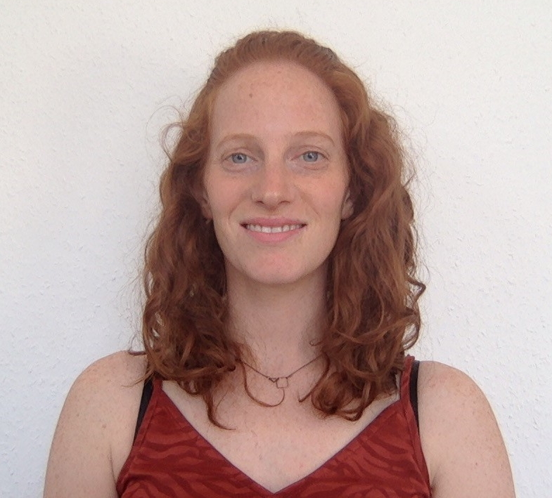
Claire Walsh is a senior Post-Doctoral Research Fellow working at UCL Department of Mechanical Engineering. After completing her PhD, at UCL investigating bubble dynamics that lead Decompression Sickness, Claire joined the interdisciplinary team of Prof. Simon Walker-Samuel and Prof. Rebecca Shipley. This project aimed to apply novel imaging techniques such as high-resolution episcopic microscopy (HREM) and Optical projection tomography (OPT) to validate the numerical simulation of CAR T-cell cancer therapy. In 2018 Claire won an MRC Skills Development Fellowship to develop digital tissue phantoms, which could be used to validate the models underpinning microstructural MRI. This work required the development of Multi-Fluorescent HREM, a 3D fluorescence microscopy technique capable of imaging individual cells within large tissue samples. In 2020 Claire became a part of an international team headed by Professor Peter Lee, which utilised the recent upgrade to the European Synchrotron Radiation Facility in Grenoble to image donated COVID-19 organs. The imaging technique developed for this project, Hierarchical Phase contrast Computed Tomography (HiP-CT), has enabled for the first time, any location within an intact human organ to be imaged with 3D histological level resolution without needed to physically section the organ. This work was recently awarded Funding from the Chan Zuckerberg Initiative for a 2.5 year proof of concept study to develop the technique further.
Talk description: Hierarchical Phase contrast Computed Tomography (HiP-CT), is a newly developed X-ray phase contrast technique which utilises the Extremely Brilliant Source at the European Synchrotron Radiation Facility. The technique is an extreme local tomography approach capable of imaging down to 1.3 um/voxel (~8um resolution) in any location within an intact human parenchymal organ (~30mm diameter) without needed to physically section. This talk will outline the technical developments, around the technique and demonstrate some of the current Biomedical applications, including application to COVID-19 lung imaging. It will also outline the future directs and challenges for the HiP-CT imaging technique and team.
David Kastner “Anatomic validation for systems neuroscience using phase-contrast Micro-CT”
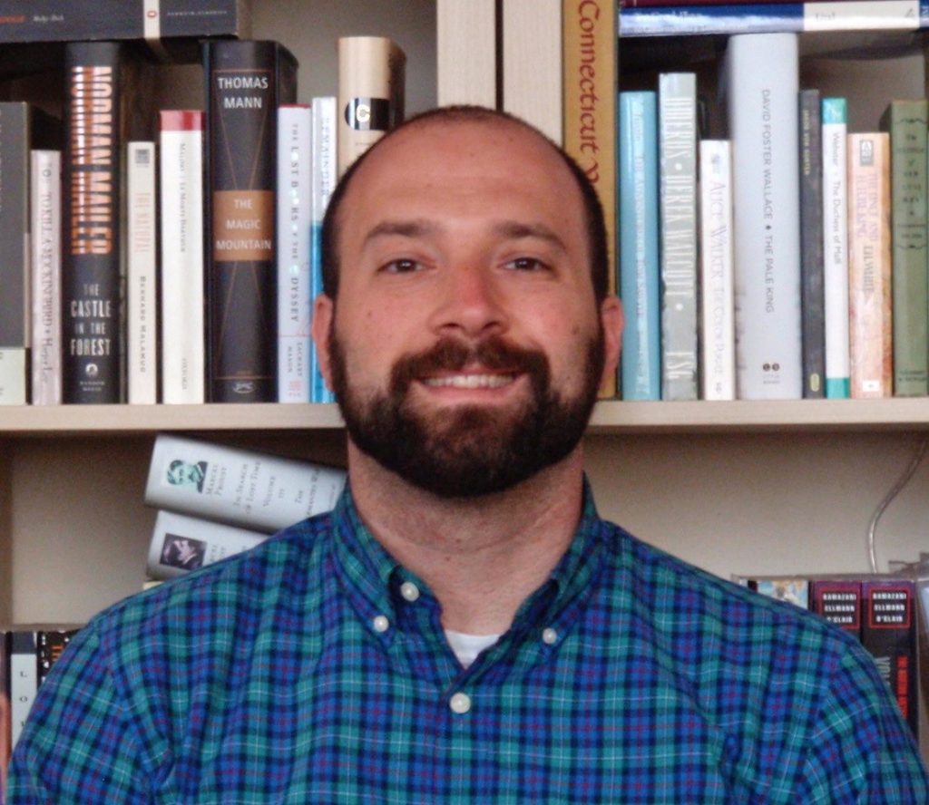
David Kastner is an Instructor in the the Department of Psychiatry and Behavioral Sciences at UCSF. He is a psychiatrist and neuroscientist. He studies the neuroscientific basis of individual variability in behavior to understand how changes in the brain lead to the extremes in behavior that occur during psychiatric illness. David earned his MD-PhD from Stanford University, working in the laboratory of Stephen Baccus and studying the function, mechanism, and theory of retinal computations. Before starting psychiatric residency at UCSF, he spent a year in Switzerland as a Fulbright Scholar working in the laboratory of Wulfram Gerstner studying synaptic levels models of consolidation and reconsolidation. During residency, David did postdoctoral research in the laboratory of Loren Frank, where he began his study of behavioral variability in the context of spatial learning and hippocampal function.
Talk description: Anatomic evaluation is an important aspect of many studies in neuroscience; however, it often lacks information about the three-dimensional structure of the brain. Micro-CT imaging provides an excellent, nondestructive method for the evaluation of brain structure, but current applications to neurophysiological or lesion studies require removal of the skull and hazardous chemicals, dehydration or embedding, limiting their scalability and utility. We have developed a protocol using eosin in combination with bone decalcification to enhance contrast in the tissue and then employ monochromatic and propagation phase-contrast micro-CT imaging to enable the imaging of brain structure with the preservation of the surrounding skull. The method allows for the quantification of brain lesions and can localize large numbers of implanted electrodes.
Adrian Wanner “Towards dynamical connectomics of cortical circuits”
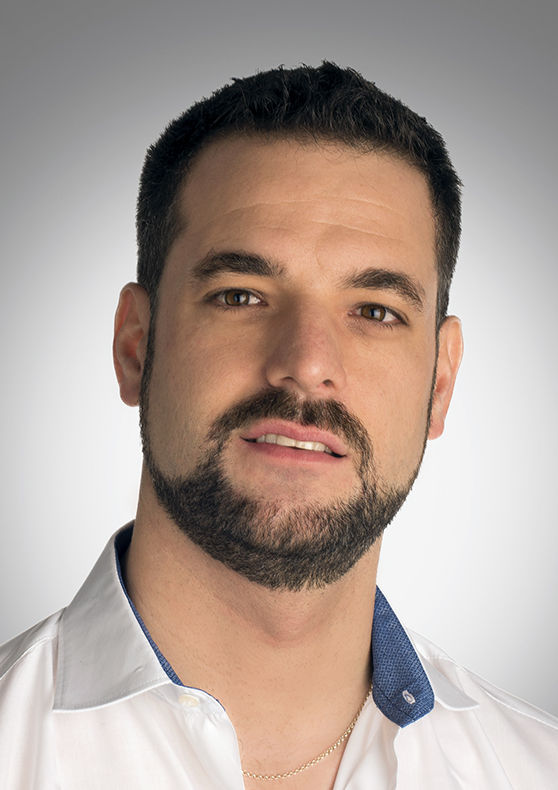
Adrian Wanner holds a MSc in Natural Sciences from ETH Zurich with majors in theoretical physics and neuroinformatics and a PhD in Neurobiology from the FMI and the University of Basel. At FMI he worked in the lab of Rainer Friedrich on combining in vivo multiphoton calcium imaging with dense, electron microscopy-based neuronal circuit reconstruction. Adrian is a CV Starr Fellow at Princeton University and investigates the structure of neuronal circuits involved in working memory of mice in collaboration with the labs of David Tank and Sebastian Seung. At the PSI in Switzerland, Adrian explores synchrotron imaging applications for neuronal circuit reconstruction.
Talk description: Dynamical connectomics produces experimentally measured blueprints of the information flow in neuronal circuits by combining dense reconstruction of neuronal circuits at synaptic resolution with functional imaging of neuronal activity. To scale this approach to cortical circuits, cubic millimeter-sized volumes of tissue have to be stained and imaged. I will present the steps we have been taking towards that goal at the Princeton Neuroscience Institute and I will discuss how to leverage connectomics with X-ray imaging.
Join the ‘Hard X-ray Imaging of Biological Soft Tissues symposium’ on Thursday 14 October 2021, 12:00-18:35 BST. Attendance is free. Register now.


 (No Ratings Yet)