Meet the people behind Volume EM community (part 1)
Posted by FocalPlane, on 21 December 2021
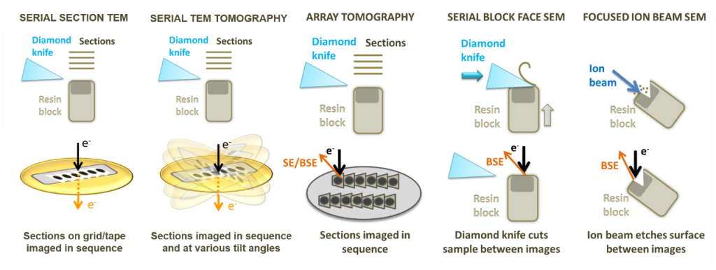
Volume Electron Microscopy or volume EM (vEM) is a relatively new term that brings together several recently developed imaging approaches that use scanning and transmission electron microscopy (SEM and TEM) to allow the interrogation of cell and tissue ultrastructure in 3D, at μm to mm volume scales and nm resolutions. In this blog series ‘volume EM’, we will post case studies using vEM techniques in different scientific fields such as cell biology, neurobiology and microbiology. As an introduction to the new blog series, FocalPlane interviewed some of the scientists involved in a new global initiative who are building an integrated community of volume Electron Microscopists and disseminating this powerful technique to researchers.

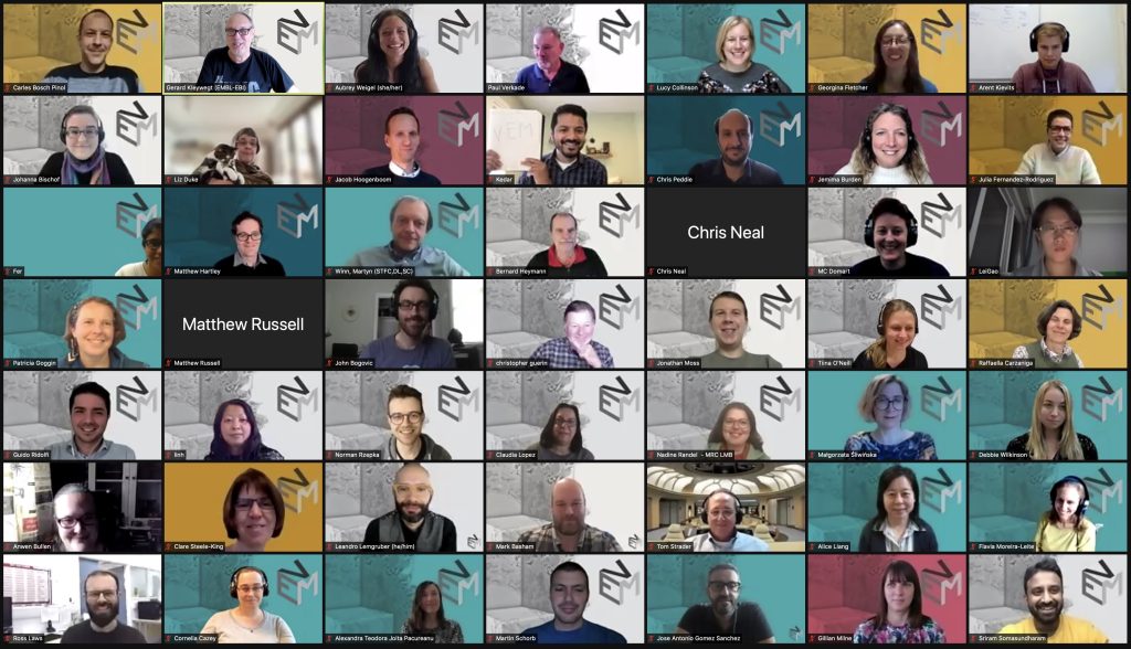
In this first post, we talked with Paul Verkade, the co-lead of this project, Anwen Bullen from the Outreach Working Group, and Chris Peddie from the Community Working Group.
Paul Verkade, Volume EM co-lead
Can you explain what the volume EM (vEM) Community Initiative is?
The vEM initiative is a community effort to bring together researchers from all disciplines who are developing and applying volume-electron microscopy techniques in their research. It is largely driven by the 6 working groups: infrastructure, community, outreach, sample preparation, training, and data.
What are the aims (goals) of the volume EM Initiative?
The vEM initiative has many goals as each Working Group has its own specific objectives. Overall, we aim to:
- Build an international community of vEM researchers.
- Reach out to and educate biomedical researchers to highlight the power of vEM for their research.
- Establish a coordinated vEM infrastructure both in the UK and internationally to benefit biomedical research.
Who is involved in this initiative?
The initiative was started by Lucy Collinson from the Crick, Gerard Kleywegt and Ardan Pathwardan from EMBL-EBI, and myself. We organised the first (in-person) vEM Town Hall meeting in 2019 with excellent support from Luigi Martino and the Wellcome Trust. From there the initiative has grown to include many of the UK vEM researchers and has now become a genuinely international effort. Whereas the first two Town Hall meetings in 2019 and 2020 attracted 50-60 participants, the third meeting in October 2021 attracted over 200 registrants from all over the world. Most of the work is done by the six working groups that have been established. Each of those has about 15-20 members that organise their activities on an entirely voluntary basis.
How did you come to be involved with the vEM Community Initiative?
My group at the University of Bristol has been doing vEM-based research for some time now (without using that term initially). Through discussions with other researchers the idea grew that we needed to bring like-minded people together and coordinate our efforts and one thing led to another. It is one of those things that you are passionate about, and so you speak up, and slowly end up completely submerged in it.
What are you doing to accomplish the vEM Initiative goals?
Most importantly we organise our Town Hall meetings. These provide an excellent opportunity for the entire community to discuss with the funding bodies what is required from both sides. It also enables us to showcase the latest scientific and technical developments in the field and to hear updates from relevant archives and related initiatives in the UK and internationally. More broadly, we are involved in organising scientific meetings, publishing vEM focussed research, developing standards and reaching out to the scientific community to offer our technologies and expertise. On that note, we are delighted that our application to organise a Gordon Research Conference (GRC) has been awarded. We will be organising the very first GRC on volume EM July 16-21, 2023 in Ventura, California, USA.
Challenges/Opportunities?
Our biggest challenge is to convince the funding bodies that it is essential to invest in this area. vEM is an umbrella term for a collection of imaging modalities that can be applied to a wide range of biological questions and are often used in concert with other techniques (such as light microscopy). Thus, making the case to the funders is more challenging than for instance in the case of the single-particle cryo-EM community a few years ago, who were basically dealing with a single type of instrument. However, having such a wide variety of researchers involved also means that we can apply the technology in many more research areas, creating impact on a much broader scale.
Which vEM discovery has struck you the most?
There have already been quite a number of scientific discoveries made using vEM but the one that really blew my mind is a very recent one. It is the demonstration of tubulated ER to Golgi carriers by Aubrey Weigel and coworkers published in Cell in 2021 (doi: 10.1016/j.cell.2021.03.035). The beauty of it is that they combine a super resolution cryo correlative microscopy approach to end up with a high-resolution volume using a special FIB-SEM. This once again shows that we critically depend on the continuous development of the technology to answer our biological questions.
Tell us something about yourself that not many people know?
Most random fact: One of the reasons I moved to the UK in 2006 was to see one of my footballing idol, Dennis Bergkamp, play for Arsenal, only for him to announce his retirement the moment I moved 😔
Anwen Bullen, Outreach Working Group
What are the aims of your WG?
The aims of the outreach working group are to understand how volume EM is being used by scientists, which scientific areas are using volume EM, and which areas could benefit for volume EM techniques, but are not currently using them. We then want to use that information to educate the scientific community about the volume EM techniques that could help their work, and about the volume EM community initiative.
Which types of people are in your WG?
Our WG has members from a range of different backgrounds, including academia, science publications and societies, commercial companies and science institutes. We have members from the UK, Europe, the USA and Australia, and we’re always happy to see new faces and new ideas!
How did you come to be involved with the vEM Community Initiative?
I was asked to speak about some of the vEM techniques at the first vEM community meeting in 2019, I thought the initiative was a great idea, and something I would really like to be involved in. One of the best parts of my day job is explaining microscopy techniques and how they can be applied to solving different scientific problems, and I was excited to get the chance to do that on a wider stage, for a set of techniques I’m really passionate about.
What are you doing to accomplish your WG’s aims?
We have been working on two main themes. To gather information from the community about how volume EM is being used, we designed a survey and ran the first version of it over the summer. Having analysed that data we are about to launch a second version of the survey to target specific areas that came out of the first survey. We are also developing outreach materials, including case studies of how volume EM is being used to solve biological problems, building a list of specialist journal reviewers for papers with volume EM topics, and publications to introduce the vEM initiative to the wider community, like this one!
Challenges/Opportunities?
Like all volunteer initiatives, we’re all working on our aims on top of our day jobs. All of our WG members have been incredibly generous with their time and hard work, but organisation across multiple time zones and around everyone’s schedules is not always easy. But our diverse backgrounds and work are a massive opportunity as well, and it really helps to have a wide variety of perspectives on microscopy and bioscience when we’re trying to design materials for a wide scientific audience.
Which vEM discovery has struck you the most?
Tricky! There have been so many beautiful pieces of vEM work. One that springs to mind is the 2017 paper by Wu et. al., where FIB-SEM was used to map contacts between endoplasmic reticulum (ER) and other membranes in neurons. It’s a great example of the strengths of vEM, a study that would be incredibly difficult to do by any other method, because of the need for both significant data volume, and electron microscopy’s ability to provide full anatomical context, and which shows how much information lies in being able to look at intracellular structures at a whole cell level. Even though (as the author’s acknowledge) they were only able to analyse a subset of neurons, their findings not only showed the complexity of ER structure and ER interactions with other organelles, but also opened up questions about what those interactions are doing.
Tell us something about yourself that not many people know?
Apart from the human and animal occupants, my house is mostly full of musical instruments. We have pianos, strings, woodwind, brass, and one pipe organ!
Chris Peddie, Community Working Group
What are the aims of your WG?
The vEM community WG aims to support everyone practically involved in the vEM ecosystem at both national and international levels. In these early stages of the initiative, we are focusing on establishing and developing networking resources, and are working to increase vEM exposure within the wider community.
Which types of people are in your WG?
The WG is a varied international collection of scientists from electron microscopy facilities, or whose work is heavily vEM orientated, be that in the microscopy itself, or in related areas such as data management. We also have a few members from the commercial sector. New members that want to be actively involved are always welcome!
How did you come to be involved with the vEM Community Initiative?
I attended both of the community meetings that preceded the launch of the initiative. These were really intriguing and demonstrated that a lot could be done to change the landscape of vEM in the UK and beyond, so I wanted to keep more closely involved in the development of the initiative. I also work with one of the ringleaders…
What are you doing to accomplish your WG’s aims?
We’ve set up a vEM mailing list, a twitter account, and a fledgling website to spread the word of the volume and hopefully serve as a central landing area for all matters vEM. We’re also looking into opportunities to increase exposure through presentations at conferences, publications, and are beginning to develop a shared resource of materials that can be used for vEM marketing. Lastly (and perhaps crucially) we recently commissioned a vEM logo for use across all platforms!

Challenges/Opportunities?
We’ve got an opportunity to establish a one-of-a-kind, linked-up resource for vEM, and create a global network of vEM experts. Keeping it relevant and maintaining engagement with microscopists as time passes will be a challenge. Managing time zones across such an international group is also very tricky!
Which vEM discovery has struck you the most?
I’m very interested in the technologies behind vEM (since in part it appeals to the part of me that really likes to take things apart and put them back together again!), and the changes that come with stepwise developments in workflows and imaging technologies, moving beyond previously show-stopping barriers in useability or application. For instance, the development of enhanced FIB SEM systems at Janelia Research Campus in order to overcome the inherent limitations of the conventional hardware, and combination with high-end ancillary techniques to provide functional context. The scale and speed at which it is now possible to collect meaningful datasets, compared to when I was a student, is astonishing.
Tell us something about yourself that not many people know?
I try to dabble a tiny bit in lunar and astrophotography, though I’m not remotely dedicated enough to sit it out during the cold nights, good whiskey notwithstanding.


 (3 votes, average: 1.00 out of 1)