Microscopy preprints – applications in cell biology
Posted by FocalPlane, on 20 May 2022
Here is a curated selection of preprints published recently. In this post, we focus specifically on preprints using microscopy tools in cell biology.
A new mechanism of fibronectin fibril assembly revealed by live imaging and super-resolution microscopy
Darshika Tomer, Cecilia Arriagada, Sudipto Munshi, Brianna E. Alexander, Brenda French, Pavan Vedula, Valentina Caorsi, Andrew House, Murat Guvendiren, Anna Kashina, Jean E. Schwarzbauer, Sophie Astrof
Actin nano-architecture of phagocytic podosomes
J. Cody Herron, Shiqiong Hu, Takashi Watanabe, Ana T. Nogueira, Bei Liu, Megan Kern, Jesse Aaron, Aaron Taylor, Michael Pablo, Teng-Leong Chew, Timothy C. Elston, Klaus M. Hahn
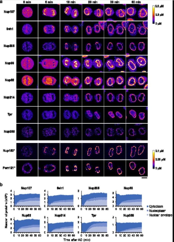
A quantitative map of nuclear pore assembly reveals two distinct mechanisms
Shotaro Otsuka, Jeremy O. B. Tempkin, Wanlu Zhang, Antonio Z. Politi, Arina Rybina, M. Julius Hossain, Moritz Kueblbeck, Andrea Callegari, Birgit Koch, Andrej Sali, Jan Ellenberg
Ultrastructure Expansion Microscopy reveals the nanoscale cellular architecture of budding and fission yeast
Kerstin Hinterndorfer, Marine. H. Laporte, Felix Mikus, Lucas Tafur Petrozzi, Clélia Bourgoint, Manoel Prouteau, Gautam Dey, Robbie Loewith, Paul Guichard, Virginie Hamel
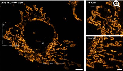
Multi-color live-cell STED nanoscopy of mitochondria with a gentle inner membrane stain
Tianyan Liu, Till Stephan, Peng Chen, Jingting Chen, Dietmar Riedel, Zhongtian Yang, Stefan Jakobs, Zhixing Chen
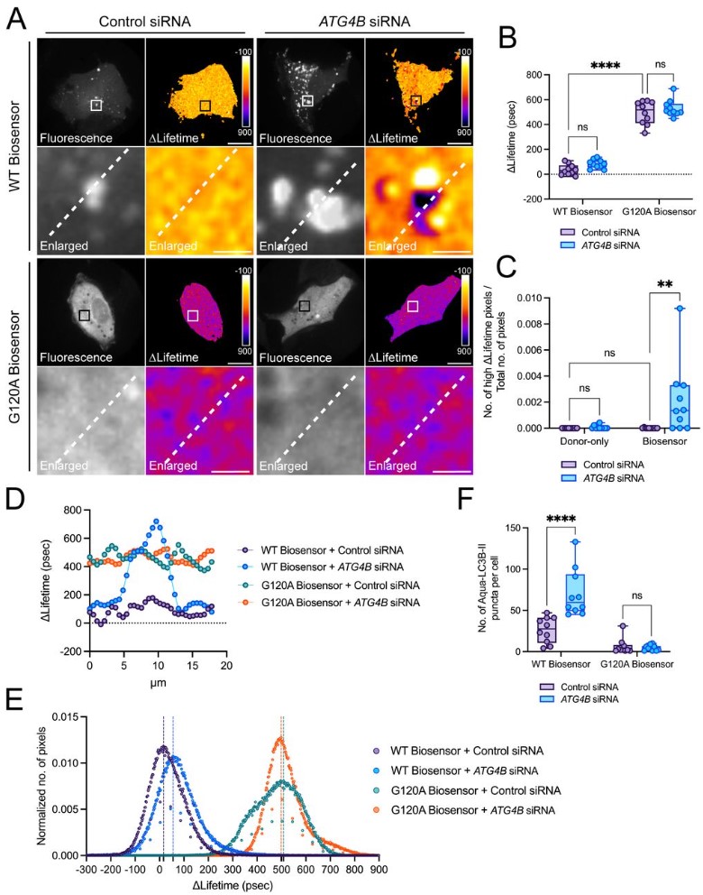
The LC3B FRET biosensor monitors the modes of action of ATG4B during autophagy in living cells
Elif Begüm Gökerküçük, Angélique Cheron, Marc Tramier, Giulia Bertolin
Intracellular connections between basal bodies promote the coordinated behavior of motile cilia
Adam W. J. Soh, Louis G. Woodhams, Anthony D. Junker, Cassidy M. Enloe, Benjamin E. Noren, Adam Harned, Christopher J. Westlake, Kedar Narayan, John S. Oakey, Philip V. Bayly, Chad G. Pearson
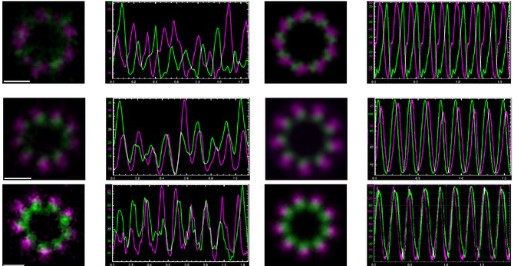
Molecular architecture of the C. elegans centriole
Alexander Woglar, Marie Pierron, Fabian Zacharias Schneider, Keshav Jha, Coralie Busso, Pierre Gönczy
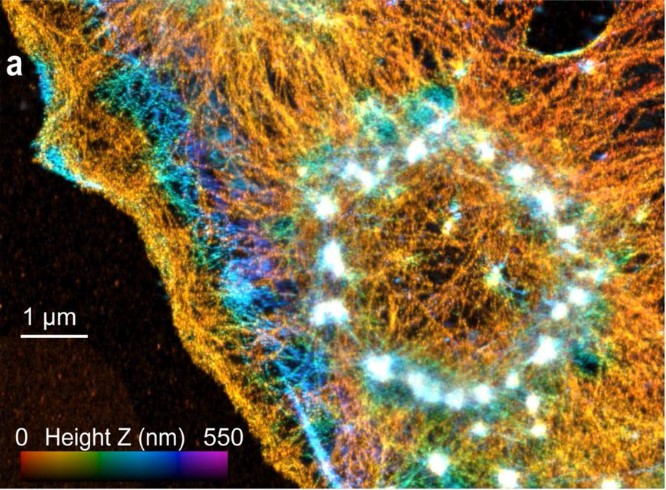
Correlative light and electron microscopy reveals fork-shaped structures at actin entry sites of focal adhesions
Karin Legerstee, Jason Sueters, Gert-Jan Kremers, Jacob P. Hoogenboom, Adriaan B Houtsmuller
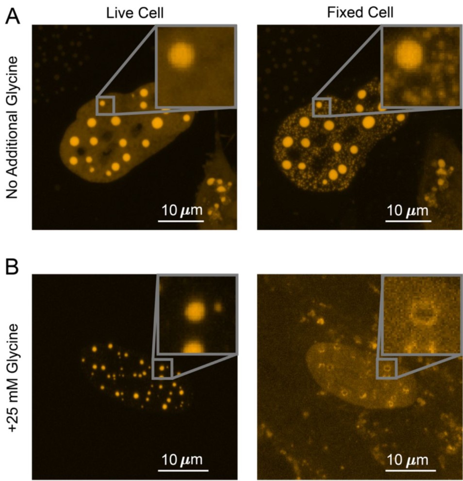
Fixation Can Change the Appearance of Phase Separation in Living Cells
Shawn Irgen-Gioro, Victoria Walling, Shasha Chong
mNeonGreen-tagged fusion proteins and nanobodies reveal localization of tropomyosin to patches, cables, and contractile actomyosin rings in live yeast cells
Tomoyuki Hatano, Tzer Chyn Lim, Ingrid Billault-Chaumartin, Anubhav Dhar, Ying Gu, Teresa Massam-Wu, Sushmitha Adhishesha, Luke Springall, William Scott, Lavanya Sivashanmugam, Masanori Mishima, Sophie G Martin, Snezhana Oliferenko, Saravanan Palani, MOHAN K BALASUBRAMANIAN
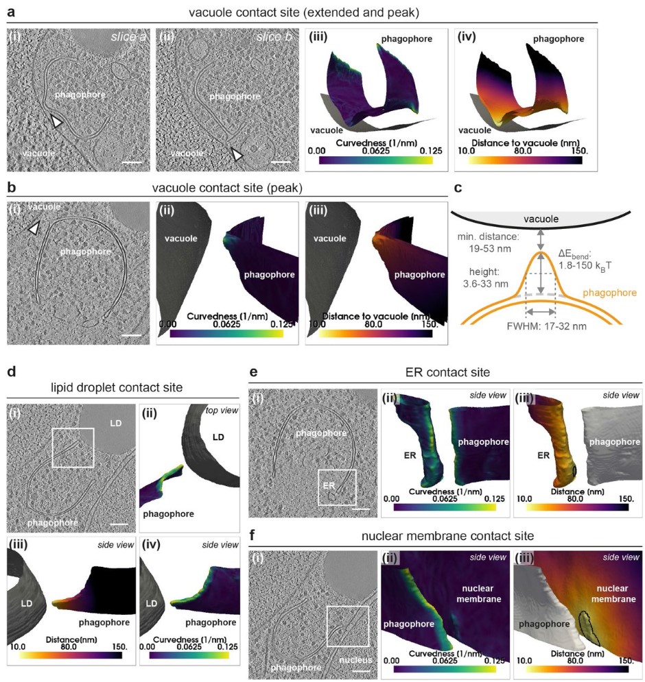
In situ structural analysis reveals membrane shape transitions during autophagosome formation
Anna Bieber, Cristina Capitanio, Philipp S. Erdmann, Fabian Fiedler, Florian Beck, Chia-Wei Lee, Delong Li, Gerhard Hummer, Brenda A. Schulman, Wolfgang Baumeister, Florian Wilfling
Spatial organization of Dectin-1 and TLR2 during synergistic crosstalk revealed by super-resolution imaging
Miao Li, Christopher Vultorius, Manisha Bethi, Yan Yu
 (No Ratings Yet)
Get involved
Create an account or log in to post your story on FocalPlane.
More posts like this

Filter by
- NewsApply
- DiscussionsApply
- How toApply
- ToolsApply
- Case studiesApply
- InterviewsApply
- JobsApply
- EducationApply
- Blog seriesApply
- Volume EMApply
- Latin American Micro..scopistsApply
- Bio-image Analysis w..ith NapariApply
- Imaging with…Apply
- Towards Global Acces..sApply
- Latin America Bioima..gingApply
- From Zero to Qupath ..HeroApply
- Asian Microscopists ..and Cell BiologistsApply
- AIC at HHMI JaneliaApply
- Deep Learning for Bi..o-image analysisApply
- GloBIAS – updates fr..om the communityApply
- WAMBIAN: West Africa.. in FocusApply
- Highlights from Euro..-BioImagingApply
- LSFM seriesApply
- DIY MicroscopyApply
- View all