EMBL Imaging Centre: Enabling imaging across scales
Posted by Virginia Pierini, on 17 June 2022
This article was written by: Shreya Ghosh, Verena Viarisio, Stephanie Alexander, Jan Ellenberg.
With its first symposium, the EMBL Imaging Centre introduced its services, aims, and missions to the scientific community, celebrated advancements in imaging, and provided a sneak-peek into the cutting-edge technologies and advanced training it will make accessible to researchers all over the world.
“There hasn’t been a meeting I have been looking forward to more in the past five years,” said Jan Ellenberg, Head of the EMBL Imaging Centre (EMBL IC) and of EMBL’s Cell Biology and Biophysics Unit, during his opening remarks to participants at the EMBL Imaging Centre Symposium: Enabling imaging across scales.
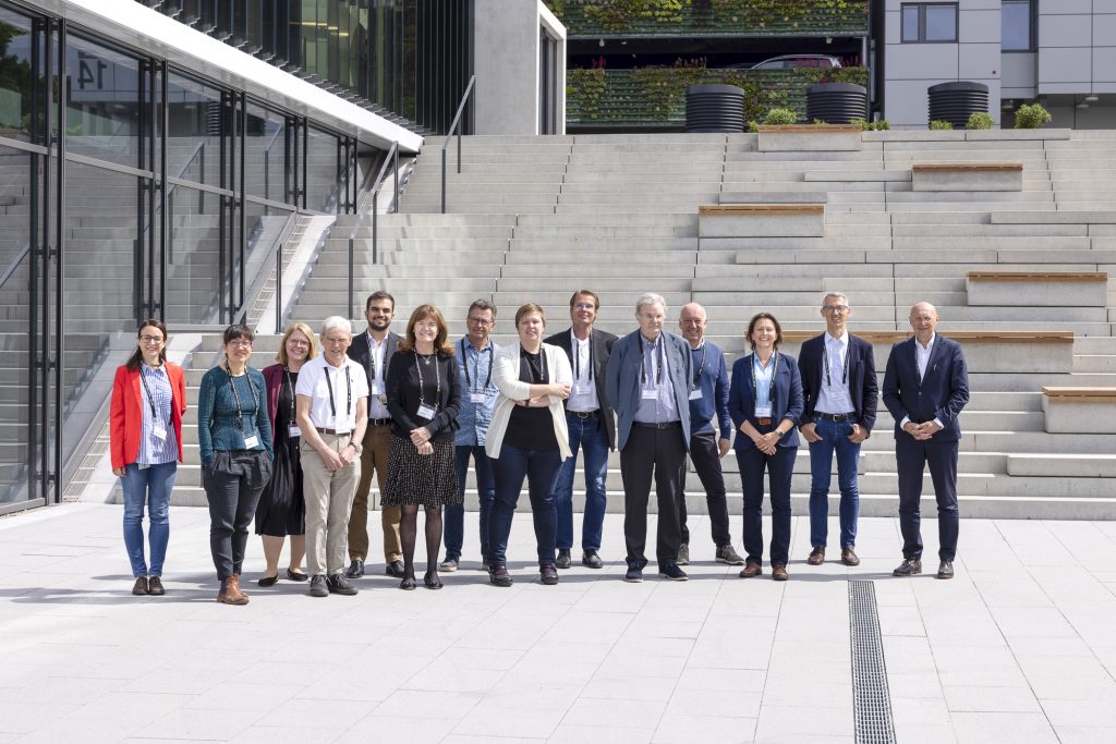
The symposium, which took place on 31 May 2022 was the first official celebration of the EMBL IC opening its doors to the scientific imaging community. For this occasion, 175 registrants and invited guests came together in person at the EMBL headquarters in Heidelberg and listened to international leaders of microscopy, including three Nobel laureates, review some of the cutting-edge technologies at the forefront of the field. In addition to witnessing a series of outstanding talks in the large auditorium of EMBL’s Advanced Training Centre, the participants had the opportunity to get to know the brand-new building that houses the EMBL Imaging Centre, which hosted the meals, poster sessions, and exhibits of the technology development partners of the EMBL IC from industry. All participants also had the chance to participate in tours of the Imaging Centre, and could connect with the speakers, the EMBL IC’s service teams, as well as each other.
In his opening address, Dr Ellenberg, who has led the planning and implementation of the EMBL IC from the conception of the idea in 2015 to the start of construction in 2018, and the beginning of service operation in late 2021, reminded the guests of the disruptive technology developments that have made microscopy into one of the most essential research tools of life science today. Once only used for anatomical or pathological studies, imaging technologies now cross biological scales in time and space, enabling researchers to resolve the structure and function of individual molecules , and do so in their natural context of entire cells and organisms.
“New technologies continue to come up and we at the EMBL Imaging Centre make them accessible to the European research community,” said Dr Ellenberg, summarising the centre’s primary mission. “We also train scientists as well as research infrastructure and facility staff in using new technologies, and promote innovation by promoting technology and workflow development with partners from academia and industry.”
Following the introductory remarks, EMBL IC’s service team leads, Simone Mattei (electron microscopy) and Timo Zimmerman (light microscopy), moderated a series of sessions in which invited speakers discussed the latest breakthroughs in imaging technology development and application.
The EMBL Imaging Centre: making the latest imaging available
In the early 2010s, EMBL groups that developed new light microscopes were increasingly flooded with requests by researchers who wanted to use the freshly minted technologies. At the same time, EMBL saw a tremendous need across Europe for the cryo-EM services and workflows that were developed in-house to tackle structural biology inside cells. Dr Jan Ellenberg, Head of Cell Biology and Biophysics Unit and Dr Christoph Müller, Head of Structural and Computational Biology Unit, acted on this by pitching for a service centre to address both of these needs and that is based on EMBL’s long-standing expertise in LM and EM: the idea for the EMBL Imaging Centre was born. In December 2021, after only two and a half years of building works and a short pilot user phase, the EMBL Imaging Centre opened its doors to users across Europe and beyond. Additionally, a permanent exhibition within the building will invite the public to get to know “The World of Molecular Biology” and how “Seeing is Understanding”.
The EMBL IC makes cutting-edge imaging technologies accessible to scientists from all around the world, from both academia and industry. The portfolio of the EMBL IC includes highest resolution electron microscopy (EM) and light microscopy (LM) technologies, correlative light and electron microscopy (CLEM) approaches, as well as technologies not yet commercially available. Supporting the open access to instruments, expert staff from two integrated service teams for LM and EM provide tailored project support during the full experimental cycle: from sample preparation to image data analysis. To disseminate knowledge about the handling of these technologies and the related workflows within the scientific community, the EMBL IC is further extending EMBL’s course and conference programme, with dedicated expert courses and regular user meetings to bring together the imaging community. Job shadowing opportunities, offered also with Global Bioimaging, targets imaging service facility staff from all around the world. Through these training multipliers, we are helping more researchers use the newest imaging technologies and workflows. In order to make new technologies available as quickly as possible, the EMBL IC works in an open innovation concept to develop technologies in close collaboration with its industry partners.
The EMBL IC Service Team is the first point of contact (ic-contact@embl.de) for all of EMBL’s external microscopy services and is happy to advise you on access routes, and to consult with you on the imaging technology options for your project. To give fair access to all users, your proposals will be quickly evaluated by independent external experts in the imaging technologies offered at the EMBL Imaging Centre. 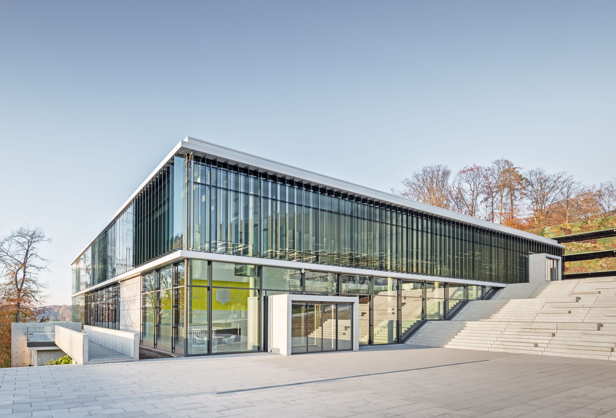
Credits: Julian Salamon / gerstner + hofmeister architects
The State of the Cryo-EM and Superresolution Microscopy Field
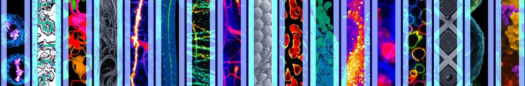
EMBL alumnus Stefan Hell, who received the Nobel Prize in Chemistry in 2014 jointly with W.E. Moerner and Eric Betzig for overcoming the resolution limit of light microscopy with his Stimlulated Emission Depletion (STED) microscope, opened the scientific sessions with a talk about ‘Fluorescence Microscopy at Molecule-Size Resolution’. Discussing the technical as well as computational challenges associated with obtaining molecular-scale resolution with fluorescence imaging methods, Dr Hell spoke about the usefulness of new techniques like MINFLUX and MINSTED. Both of these techniques minimise the photobleaching effects of previous methods, and together, they push the resolution range of fluorescence microscopy to the 1-3 nm scale, at which point, resolution is no longer limited by the precision of the techniques, but by the size of the fluorophores themselves. Dr Hell illustrated the application of these methods with beautifully resolved images of nuclear pore complexes (a collaboration with researchers at EMBL), mitochondrial proteins, and lipid molecules moving within bilayers.
One of the techniques that has revolutionised the field of structural biology in recent years is cryogenic electron microscopy (cryo-EM), which allows researchers to visualise the molecular building blocks of life at an unprecedented level of detail. Richard Henderson, who was jointly awarded the Nobel Prize in Chemistry in 2017 with Joachim Frank and EMBL alumnus Jacques Dubochet for developing this technique, aptly titled his talk ‘Future Trends in cryo-EM’. Walking the audience through some of the major milestones in the application of cryo-EM technologies in the last few decades, Dr Henderson commented on the exponential growth of protein data bank (PDB) entries that has resulted from the proliferation of this technology. He emphasised the need for more affordable cryo-EM techniques to make cryo-EM more widely accessible to non-specialists, and provided a technical roadmap for how this can be achieved. In addition, Dr Henderson predicted that future major hardware developments in cryo-EM could consist of several other hardware components, including better phase plates, very low temperature cold-stages, and chromatic aberration correctors. He also highlighted the need for more advanced computational techniques, better ways of freezing samples, and robustly making thin sections from vitrified tissue.
The aspect of sample preparation was also a major focus of Bridget Olivia Carragher from Columbia University Irving Medical Center, New York. In her talk ‘cryo-EM Automation: Better, Faster, Cheaper, and Smarter’, she emphasised that while electron microscopy is on a fantastic exponential rise, the community needs to keep innovating, especially on improving the sample preparation step in the cryo-EM workflows. According to her, the key to speedier and thus cheaper data collection would be to reliably hit just the sweet spot for the correct ice thickness and to optimise grid preparation. To this end, she introduced the Spotiton project, which resulted in the development of a self-blotting nanowire grid, overcoming many of the issues that arise from the interaction of particles with the air-water interface.
In the symposium’s final talk ‘Recent Advances in Correlative Microscopy and Super-Resolution Imaging of Coronavirus Infection’, Nobel laureate W.E. Moerner highlighted the power of correlating electron and light microscopy techniques under cryogenic conditions, using the example of a project that mapped the biochemistry of coronavirus replication within the host cell. He also spoke about the challenges of annotating hidden proteins and structures in a cryo-EM images, and approaches to overcoming this challenge. Dr Moerner emphasised how the two recent revolutions in spatial resolution – super-resolution optical microscopy and cryo-EM – have made such investigations possible, and how the combination of both technologies could result in insights beyond the abilities of each technology individually to achieve. Together they could revolutionise our understanding of fundamental dynamic molecular processes, such as cellular infection.
Molecules to Organelles to Embryos
The remaining talks focused on some groundbreaking applications of novel imaging technologies. Suliana Manley, Swiss Federal Institute of Technology (EPFL), Lausanne, Switzerland, spoke about ‘Biophysical mysteries and insights into organelle structure and dynamics’. Discussing how structure often determines function when it comes to organelles, she spoke about the importance of leveraging both automation and parallelisation to understand dynamic structural variability in organelles. She described her own successes at achieving large fields of view using a combination of STORM, DNA-PAINT, and iSIM technologies, which allowed her to observe mitochondrial dynamics at a high spatial and temporal resolution. This, in turn, allowed her team to figure out the physiological significance of localised variations in mitochondrial fission dynamics. She also proposed that the future of microscopy lies in ‘intelligent’ systems which would be adaptive, shifting temporal and spatial resolution to match dynamic events happening in the biological system under observation.
In his talk titled ‘Structural Biology in situ or The Power of Seeing the Whole Picture’, Wolfgang Baumeister, Max Planck Institute of Biochemistry, Germany, discussed how while initially only really thin and small objects, such as viruses, could be imaged by cryo-electron tomography (cryo-ET), this is now no longer the case. For example, techniques such as focused ion beam milling allows the creation of thin and compression-free windows into cells. Dr Baumeister illustrated the power of advanced tomography in structural biology through results from his group’s work carrying out in situ studies of Chlamydomonas nuclear proteasomes. For realising the full potential of cryo-ET, he suggested that it would be important to integrate the label-free structural power of cryo-electron microscopy with the molecular specificity of cryo-fluorescence microscopy, as well as to come up with automated procedures for data mining and ways to integrate complementary information into the analysis.
Gaia Pigino, Structural Biology Center at Human Technopole, Italy, delivered a talk on the ‘Structural cell biology of cilia and eukaryotic flagella’. First stating the importance of cilia to human physiology and the many pathologies that arise from ciliary dysfunction – ciliopathies, she went on to explain the fascinating system of anterograde and retrograde cargo trains carried by two different motor proteins, which are necessary for ciliary assembly. Using correlative light and electron microscopy (CLEM), her team was able to discover that anterograde and retrograde trains use different tracks, and use this information to subsequently figure out the 3D architecture of intraflagellar transport trains using cryo-ET. Dr Pigino’s group subsequently studied the assembly of the anterograde train at the ciliary base using cryo-FEB-SEM, and plans to use expansion fluorescence microscopy to further investigate these processes in the future.
Moving from the subcellular to the organismal scale, Kate McDole, MRC Laboratory of Molecular Biology, UK, spoke about ‘Imaging and reconstructing early mouse development using light-sheet microscopy’. Discussing her own ‘lockdown project’ – creating a mouse SiMView microscope, specifically tailored for the requirements of the mammalian embryo – Dr McDole stressed how the advent of advanced imaging technologies has resulted in a modern ‘renaissance’ for the field of developmental biology. Since the optical properties and scale of tissues change as an embryo grows, real-time adaptive imaging is needed to keep track of the embryo in space and time, which the SiMView set-up achieves. Additionally, it allows the researcher to visualise multiple reporter combinations, follow single-cell behaviours, and track cell lineages across long developmental time scales, while precisely manipulating and perturbing developmental events as they happen. Dr McDole also discussed the rising computational complexity associated with large datasets and the importance of open source community software, public image data archives, and new data analysis approaches to this process.
The Road Ahead
Before the audience moved on to an evening of networking and social activities, Jan Ellenberg thanked the speakers and the EMBL IC technology partners – Abberior Instruments, Carl Zeiss Microscopy, Leica Microsystems and Thermo Fischer Scientific – who also provided demonstrations of their instruments in the EMBL IC to the participants. In his closing remarks, Dr Ellenberg marvelled at the outstanding achievements in the field of microscopy that were discussed at the symposium. “Something that was unthinkable only some years ago, the determination of atomic structure of macromolecules inside cells, is now routine,” he said. He closed the session by reminding the participants of the challenges ahead: “One of the biggest challenges will be to get comprehensive views of whole cells and multicellular organisms. In order to do so, we need to develop new technologies, some of which we have learned about today, and we need to make them available to the life science community quickly to move the field forward.” One of the platforms to do so is the EMBL IC, which is also part of the pan-European research infrastructures Euro-BioImaging and Instruct.
Many of the invited speakers lauded the EMBL IC and what it represents for the life sciences community. “It’s a great pleasure to help celebrate the opening of the new imaging centre,” said W.E. Moerner. “It couldn’t be more timely and it will be second to none,” agreed Stefan Hell.
If you are interested in learning more about the EMBL Imaging Centre and accessing routes to apply for project support, please visit the EMBL Imaging Centre homepage and contact the service team.
 (No Ratings Yet)
Get involved
Create an account or log in to post your story on FocalPlane.
More posts like this

Filter by
- NewsApply
- DiscussionsApply
- How toApply
- ToolsApply
- Case studiesApply
- InterviewsApply
- JobsApply
- EducationApply
- Blog seriesApply
- AIC at HHMI JaneliaApply
- Deep Learning for Bi..o-image analysisApply
- GloBIAS – updates fr..om the communityApply
- WAMBIAN: West Africa.. in FocusApply
- Volume EMApply
- Latin American Micro..scopistsApply
- Bio-image Analysis w..ith NapariApply
- Imaging with…Apply
- Towards Global Acces..sApply
- Latin America Bioima..gingApply
- From Zero to Qupath ..HeroApply
- Asian Microscopists ..and Cell BiologistsApply
- Highlights from Euro..-BioImagingApply
- LSFM seriesApply
- DIY MicroscopyApply
- View all