Microscopy preprints – applications in cell biology
Posted by FocalPlane, on 1 July 2022
Here is a curated selection of preprints published recently. In this post, we focus specifically on preprints using microscopy tools in cell biology.
Superresolution microscopy reveals partial preassembly and subsequent bending of the clathrin coat during endocytosis
Markus Mund, Aline Tschanz, Yu-Le Wu, Felix Frey, Johanna L. Mehl, Marko Kaksonen, Ori Avinoam, Ulrich S. Schwarz, Jonas Ries

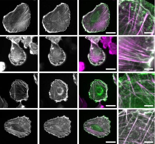
IntAct: a non-disruptive internal tagging strategy to study actin isoform organization and function
M.C. van Zwam, W. Bosman, W. van Straaten, S. Weijers, E. Seta, B. Joosten, K. van den Dries
NISNet3D: Three-Dimensional Nuclear Synthesis and Instance Segmentation for Fluorescence Microscopy Images
Liming Wu, Alain Chen, Paul Salama, Kenneth Dunn, Edward Delp

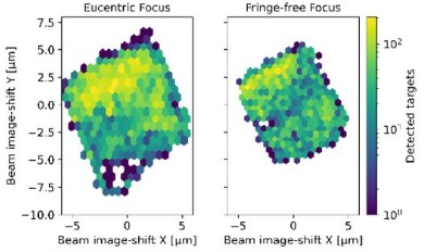
Defocus Corrected Large Area Cryo-EM (DeCo-LACE) for Label-Free Detection of Molecules across Entire Cell Sections
Johannes Elferich, Giulia Schiroli, David Scadden, Nikolaus Grigorieff
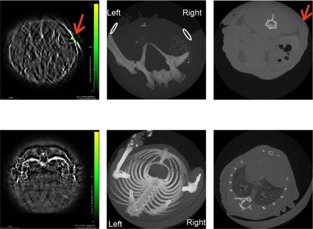
Computed tomography lacks sensitivity to image gold labelled mesenchymal stromal cells in vivo as evidenced by multispectral optoacoustic tomography
Alejandra Hernandez Pichardo, James Littlewood, Arthur Taylor, Bettina Wilm, Raphaël Lévy, Patricia Murray
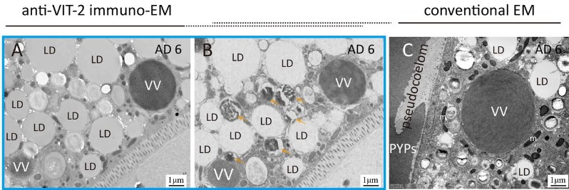
Immuno-electron microscopy localizes Caenorhabditis elegans vitellogenins along the classic exocytosis route
Chao Zhai, Nan Zhang, Xi-Xia Li, Xue-Ke Tan, Fei Sun, Meng-Qiu Dong
DNA packaging via hierarchical chromatin structures revealed by live-cell 3D imaging
Yang Zheng, Sen Ye, Shumin Li, Cuifang Liu, Shihang Luo, Yanqin Chen, Yunsheng Li, Lingyi Huang, Shan Deng, Ping Chen, Yongdeng Zhang, Wei Ji, Ruibang Luo, Guohong Li, Dan Yang
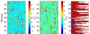
Granger-causal inference of the lamellipodial actin regulator hierarchy by live cell imaging without perturbation
Jungsik Noh, Tadamoto Isogai, Joseph Chi, Kushal Bhatt, Gaudenz Danuser
Protein nanobarcodes enable single-step multiplexed fluorescence imaging
Daniëlle de Jong-Bolm, Mohsen Sadeghi, Guobin Bao, Gabriele Klaehn, Merle Hoff, Lucas Mittelmeier, F. Buket Basmanav, Felipe Opazo, Frank Noé, Silvio O. Rizzoli
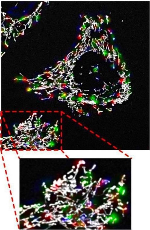
Three-dimensional mitochondrial fission, fusion and depolarisation event location prediction for a high throughput analysis of fluorescence microscopy images
James Garrett de Villiers, Rensu Petrus Theart
Three-dimensional structure of kinetochore-fibers in human mitotic spindles
Robert Kiewisz, Gunar Fabig, William Conway, Daniel Baum, Daniel Needleman, Thomas Müller-Reichert
 (No Ratings Yet)
Get involved
Create an account or log in to post your story on FocalPlane.
More posts like this

Filter by
- NewsApply
- DiscussionsApply
- How toApply
- ToolsApply
- Case studiesApply
- InterviewsApply
- JobsApply
- EducationApply
- Blog seriesApply
- Volume EMApply
- Latin American Micro..scopistsApply
- Bio-image Analysis w..ith NapariApply
- Imaging with…Apply
- Towards Global Acces..sApply
- Latin America Bioima..gingApply
- From Zero to Qupath ..HeroApply
- Asian Microscopists ..and Cell BiologistsApply
- AIC at HHMI JaneliaApply
- Deep Learning for Bi..o-image analysisApply
- GloBIAS – updates fr..om the communityApply
- WAMBIAN: West Africa.. in FocusApply
- Highlights from Euro..-BioImagingApply
- LSFM seriesApply
- DIY MicroscopyApply
- View all