Microscopy preprints – new tool and techniques in imaging and image analysis
Posted by FocalPlane, on 29 July 2022
Here is a curated selection of preprints published recently. In this post, we focus specifically on preprints on new tools and techniques in imaging and image analysis.
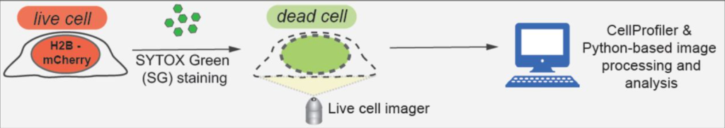
Live-cell imaging and mathematical analysis of the “community effect” in apoptosis
Diane Coursier, David Coulette, Hélène Leman, Emmanuel Grenier, Gabriel Ichim
Fluorogenic U-rich internal loop (FLURIL) tagging with bPNA enables intracellular RNA and DNA tracking
Yufeng Liang, Sydney Willey, Yu-Chieh Chung, Yi-Meng Lo, Shiqin Miao, Sarah Rundell, Li-Chun Tu, Dennis Bong
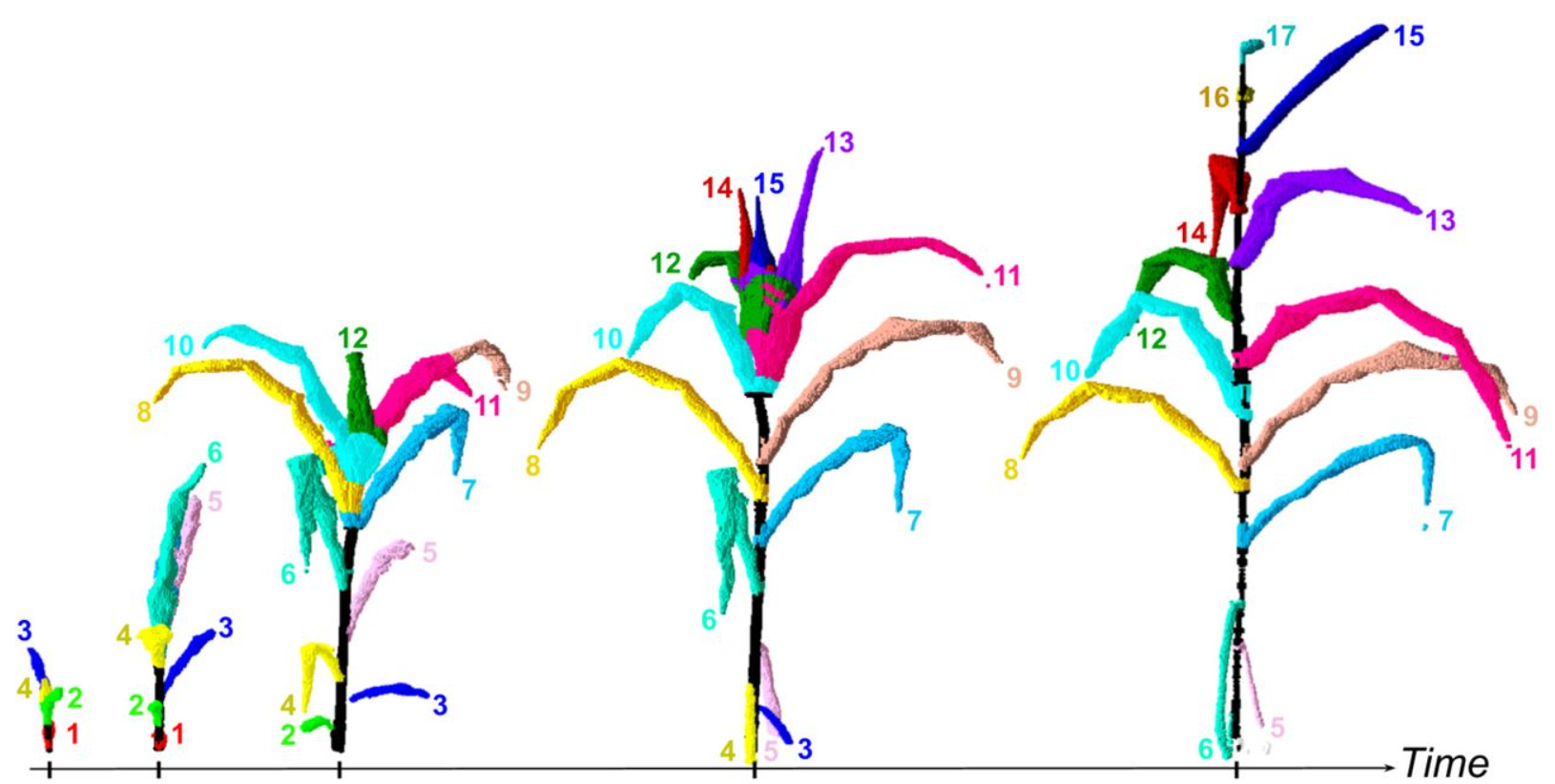
PhenoTrack3D: an automatic high-throughput phenotyping pipeline to track maize organs over time
Benoit Daviet, Romain Fernandez, Llorenç Cabrera-Bosquet, Christophe Pradal, Christian Fournier
A self-blinking DNA probe for live-cell superresolution 3D imaging of hierarchical chromatin structures
Yang Zheng, Sen Ye, Shumin Li, Cuifang Liu, Shihang Luo, Yanqin Chen, Yunsheng Li, Lingyi Huang, Shan Deng, Ping Chen, Yongdeng Zhang, Wei Ji, Ruibang Luo, Guohong Li, Dan Yang
Three-dimensional structured illumination microscopy with enhanced axial resolution
Xuesong Li, Yicong Wu, Yijun Su, Ivan Rey-Suarez, Claudia Matthaeus, Taylor B. Updegrove, Zhuang Wei, Lixia Zhang, Hideki Sasaki, Yue Li, Min Guo, John P. Giannini, Harshad D. Vishwasrao, Jiji Chen, Shih-Jong J. Lee, Lin Shao, Huafeng Liu, Kumaran S. Ramamurthi, Justin W. Taraska, Arpita Upadhyaya, Patrick La Riviere, Hari Shroff
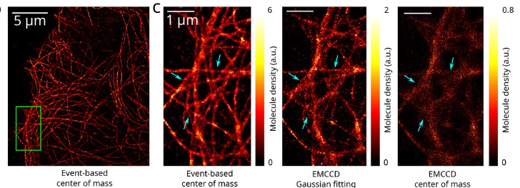
Event-based vision sensor enables fast and dense single-molecule localization microscopy
Clément Cabriel, Christian G. Specht, Ignacio Izeddin
ALIBY: ALFA Nanobody-Based Toolkit for Imaging and Biochemistry in Yeast
Dipayan Akhuli, Anubhav Dhar, Aileen Sara Viji, Bindu Bhojappa, Saravanan Palani
Untrained, physics-informed neural networks for structured illumination microscopy
Zachary Burns, Zhaowei Liu
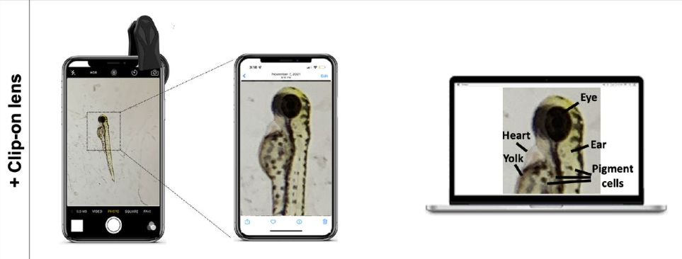
A low-cost smartphone fluorescence microscope for research, life science education, and STEM outreach
Madison A Schaefer, Heather N Nelson, John L Butrum, James R Gronseth, Jacob H Hines
Single-molecule localization microscopy reveals the ultrastructural root constitution of distal appendages in expanded mammalian centrioles
Ting-Jui Ben Chang, Jimmy Ching-Cheng Hsu, T. Tony Yang
u-track 3D: measuring and interrogating dense particle dynamics in three dimensions
Philippe Roudot, Wesley R. Legant, Qiongjing Zou, Kevin M. Dean, Tadamoto Isogai, Erik S. Welf, Ana F. David, Daniel W. Gerlich, Reto Fiolka, Eric Betzig, Gaudenz Danuser
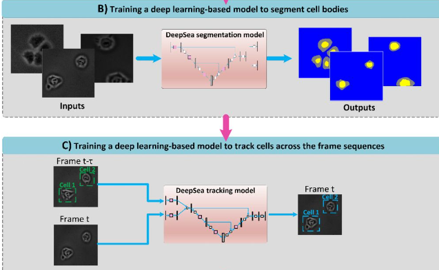
DeepSea: An efficient deep learning model for single-cell segmentation and tracking of time-lapse microscopy images
Abolfazl Zargari, Gerrald A. Lodewijk, Najmeh Mashhadi, Nathan Cook, Celine W. Neudorf, Kimiasadat Araghbidikashani, Stefany Rubio, Eva Hrabeta-Robinson, Angela N. Brooks, Lindsay Hinck, S. Ali Shariati
High-plex Multiomic Analysis in FFPE at Subcellular Level by Spatial Molecular Imaging
Shanshan He, Ruchir Bhatt, Carl Brown, Emily A. Brown, Derek L. Buhr, Kan Chantranuvatana, Patrick Danaher, Dwayne Dunaway, Ryan G. Garrison, Gary Geiss, Mark T. Gregory, Margaret L. Hoang, Rustem Khafizov, Emily E. Killingbeck, Dae Kim, Tae Kyung Kim, Youngmi Kim, Andrew Klock, Mithra Korukonda, Alecksandr Kutchma, Zachary R. Lewis, Yan Liang, Jeffrey S. Nelson, Giang T. Ong, Evan P. Perillo, Joseph C. Phan, Tien Phan-Everson, Erin Piazza, Tushar Rane, Zachary Reitz, Michael Rhodes, Alyssa Rosenbloom, David Ross, Hiromi Sato, Aster W. Wardhani, Corey A. Williams-Wietzikoski, Lidan Wu, Joseph M. Beechem
A FRET biosensor, SMART, monitors necroptosis in renal tubular epithelial cells in a cisplatin-induced kidney injury model
Shin Murai, Kanako Takakura, Kenta Sumiyama, Kenta Moriwaki, Kenta Terai, Sachiko Komazawa-Sakon, Yoshifumi Yamaguchi, Tetuo Mikami, Kimi Araki, Masaki Ohmuraya, Michiyuki Matsuda, Hiroyasu Nakano
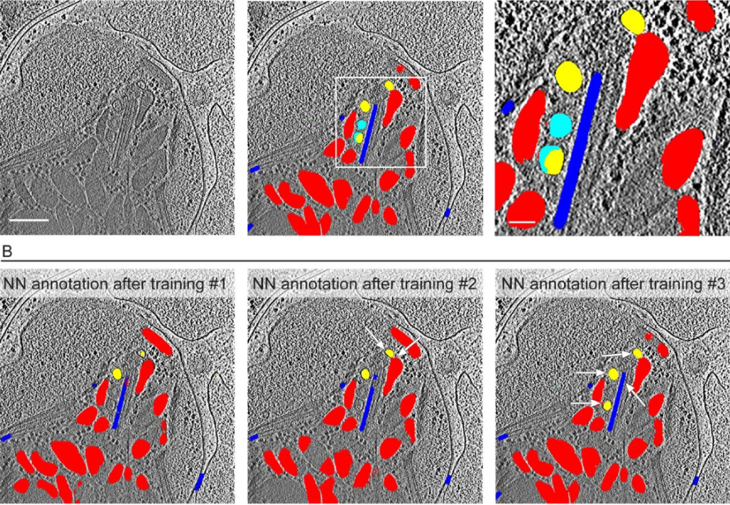
Coupling cryo-electron tomography with mixed-scale dense neural networks reveals re-organization of the invasion machinery of Toxoplasma gondii upon ionophore-stimulation
Li-av Segev-Zarko, Peter D. Dahlberg, Stella Y. Sun, Daniël M. Pelt, Chi Yong Kim, Elizabeth S. Egan, James A. Sethian, Wah Chiu, John C. Boothroyd
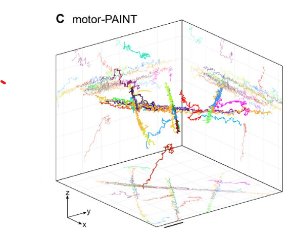
Direct observation of motor protein stepping in living cells using MINFLUX
Takahiro Deguchi, Malina K. Iwanski, Eva-Maria Schentarra, Christopher Heidebrecht, Lisa Schmidt, Jennifer Heck, Tobias Weihs, Sebastian Schnorrenberg, Philipp Hoess, Sheng Liu, Veronika Chevyreva, Kyung-Min Noh, Lukas C. Kapitein, Jonas Ries
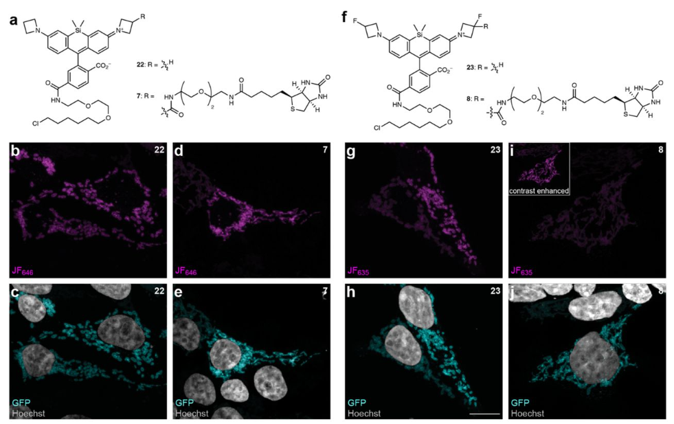
Multifunctional fluorophores for live-cell imaging and affinity capture of proteins
Pratik Kumar, Jason D. Vevea, Edwin R. Chapman, Luke D. Lavis
 (No Ratings Yet)
Get involved
Create an account or log in to post your story on FocalPlane.
More posts like this

Filter by
- NewsApply
- DiscussionsApply
- How toApply
- ToolsApply
- Case studiesApply
- InterviewsApply
- JobsApply
- EducationApply
- Blog seriesApply
- Asian Microscopists ..and Cell BiologistsApply
- AIC at HHMI JaneliaApply
- Deep Learning for Bi..o-image analysisApply
- GloBIAS – updates fr..om the communityApply
- WAMBIAN: West Africa.. in FocusApply
- Volume EMApply
- Latin American Micro..scopistsApply
- Bio-image Analysis w..ith NapariApply
- Imaging with…Apply
- Towards Global Acces..sApply
- Latin America Bioima..gingApply
- From Zero to Qupath ..HeroApply
- Highlights from Euro..-BioImagingApply
- LSFM seriesApply
- DIY MicroscopyApply
- View all