An interview with Francisco Barrantes
Posted by Mariana De Niz, on 20 September 2022

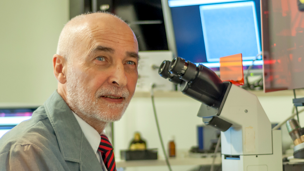
MiniBio: Prof. Francisco Barrantes got his MD and Ph.D. at the University of Buenos Aires. He is a former joint head of Membrane Biophysics Unit (1978-1983) with Erwin Neher and Bert Sakmann (Nobel awardees Med. & Physiol. 1999), at Max-Planck Institute for Biophysical Chemistry in Göttingen, Germany. During his career, he established collaborations in Germany with Derek Marsh, Tom Jovin, Peter Zinghsheim and Joachim Frank (Nobel awardee in Chemistry 2017). He is also a former head of the Scientific & Technol. Research Council of Argentina, Bahia Blanca, and a Chairholder for UNESCO (Chair of Molecular Neurobiology & Biophysics). Subsequent sabbatical periods in Göttingen led to a long-lasting collaboration with Stefan Hell (Nobel awardee in Chemistry 2014) on superresolution optical microscopy.
During his career, he obtained multiple awards, including a TWAS award in Biology; Fellow, Guggenheim Mem. Foundation; Human Frontier Science Programme Fellow; Royal Society, UK; De Robertis Medal; Alexander von Humboldt Foundation award; Premio União Latina, Portugal; Konex Award; Sarojini Damodaran Intl. Trust, India; Consecration Medal, Acad. Sci. Argentina. Gregorio Weber award, Biophys. Soc. He is currently a Member of the National Academy of Medicine and the National Academy of Sciences, Argentina; the Brazilian Academy of Science; the European Academy of Sciences; the Indian Science Academy, and the Latin American Academy of Science.
What inspired you to become a scientist?
I recall playing with anilines at my grandparent´s house at the age of about 5 and during all my primary school days I used to tell everyone I wanted to be a chemist. One of my uncles worked at a large optician’s, where I saw microscopes for the first time. I was intrigued! While at high school I attended a clinical biochemistry lab where I became acquainted with the use of a microscope and imaged blood cells. When I went to medical school at the University of Buenos Aires, I debated whether I wanted to become a basic scientist or a psychiatrist. My first-year histology examination was the turning point. My decision was made.
You have a career-long involvement in lipid and cell biology, and microscopy. Can you tell us a bit about what inspired you to choose these paths?
In my view the topics one addresses along a scientific career are 90% a combination of opportunities and serendipity and 10% creative inspiration. While still a graduate student I wrote a review article on membranes, which had to be submitted anonimously. My entry won and the prize was handed to me by Nobel awardee Luis Leloir. This early gratifying feedback must have had quite an impact on my future choice of topics, among which membranes have always had a priviledged ranking. My supervisor, cell biologist and neurobiologist Eduardo De Robertis, gave me ample freedom to choose the specific subject matter of my thesis work. He was very much interested in a specialized type of subcellular organelle, the synapse. I started exploring the molecular constituents of the cholinergic synapse as an undergraduate and continue to be very much interested in this area. I work on the nicotinic receptor, the protein that transduces the chemical signal conveyed by the neurotransmitter acetylcholine into an excitatory signal in a brain neuron or into a contraction of the skeletal muscle cell. Currently in our group we look at this molecule with a variety of microscopes to learn about their “social behavior”, metaphorically speaking, that is exploring the way individual molecules associate into the supramolecular assembly we call synapse.
Can you tell us a bit about what you have found uniquely positive about becoming a researcher in Argentina, from your education years?
As a medical student, when I took the Histology exam during my first year, Professor Eduardo De Robertis asked me whether I would like to become one of his teaching assistants. Of course, I gladly accepted. I spent a decade with him, during the period that was probably the “golden era” of the Institute under his direction. De Robertis had gathered a unique group of talented neuroanatomists, electron microscopists, biochemists, cytologists and embryologists, and the place was a mecca for young scientists, several of them foreigners. De Robertis chose me to learn fluorescence spectroscopy alongside Gregorio Weber when Gregorio returned to Argentina after several decades abroad. Gregorio became mi guru in the fluorescence field, and guided me in key steps of my future career. He suggested I go to the Max-Planck-Institute for Biophyical Chemistry in Germany, which I did… and stayed for 10 years…
Once you chose microscopy as a profession or main discipline, can you expand more about how your career has progressed in this line?
I do not consider myself a microscopist but a neuroscientist at large. I taught histology for more than a decade at the University of Buenos Aires, during which period I was also trained as an electron microscopist. I continued using this technique in Germany. Back in Argentina I made more frequent use of light microscopy. Royal Society/Human Frontier fellowships enabled me to spend a sabbatical visit (1990-1991) at the LMB in Cambridge, U.K. with one of the pioneers of cryo-electron microscopy, Nigel Unwin. An Alexander von Humboldt research award in 1999 gave me the possibility of spending short sabbatical periods at the Max-Planck in Göttingen, where I met the young head of the NanoBiophotonics Department, Stefan Hell. This coincided with a revolution in optical microscopy, with the advent of superresolution microscopy and the breaking of the diffraction limit.
Did you have many opportunities to interact with other Latin American groups, outside of Argentina?
Yes. Upon returning from my decade-long stay in Germany in 1983, I was frequently invited to teach at scientific courses at the Center for Scientific Studies (CECs) of Santiago, Chile, headed by theoretical physicist Claudio Bunster and biochemist Ramón Latorre. In this uniquely endowed institution I met many other Latin American scientists. I had joint research projects and published together with Ramón and with Nibaldo Inestrosa at the Pontifical Catholic University of Chile, another prestigious institution. I also interacted extensively with colleagues in Uruguay; with Federico Dajas at the Clemente Estable Institute (where De Robertis had worked for several years during his exile) and with Gonzalo Ferreira at Universidad de la República in Montevideo. Ramón, Nibaldo, Federico, and Gonzalo, together with several other professors, frequently participated as visiting teaching staff at postgraduate courses I organized as head of the UNESCO Chair of Biophysics & Molecular Neurobiology in Bahía Blanca. I was also often invited to teach at the Chagas Institute of Biophysics of the Federal University of Rio de Janeiro by Leopoldo De Meis, and more recently by Wanderley De Souza.

Have you ever faced any specific challenges as an Argentinian researcher, working abroad?
Of course there are always challenges of one sort or another when one leaves one’s home-country to work abroad, but in all my years of working abroad I don’t think I ever faced challenges of a serious nature. Intellectual challenges were always stimulating and enjoyable. My wife and I lived in Germany for a decade. We were aware that becoming integrated required a positive and proactive attitude, and this proved the key to becoming rapidly integrated and making friends – friendships which have lasted ever since. My children were born in Göttingen and attended kindergarden and part of their primary school there. The entire family spent sabbatical periods at Stony Brook in New York, at the Laboratory of Molecular Biology of the Medical Research Council in Cambridge, U.K., the children attending school and my wife engaged in voluntary work at the university museum art restoration facility. . My eldest daughter, of secondary school age, did some voluntary work at Addenbrooks Hospital. I had a very tight schedule during our decade at the Max-Planck-Institute in Göttingen, travelling to innumerable meetings and visits abroad. Under none of these circumstance did I or my family experience insurmountable challenges.
Who are your scientific role models (both Argentinian and foreign)?
I have been very fortunate during my scientific career, meeting many talented scientists and a few exceptional ones. The closest approximation to a genius that I’ve met was Manfred Eigen. Gregorio Weber and Eduardo De Robertis remain my role models.
What is your opinion on gender balance in Argentina, given current initiatives in the country to address this important issue. How has this impacted your career?
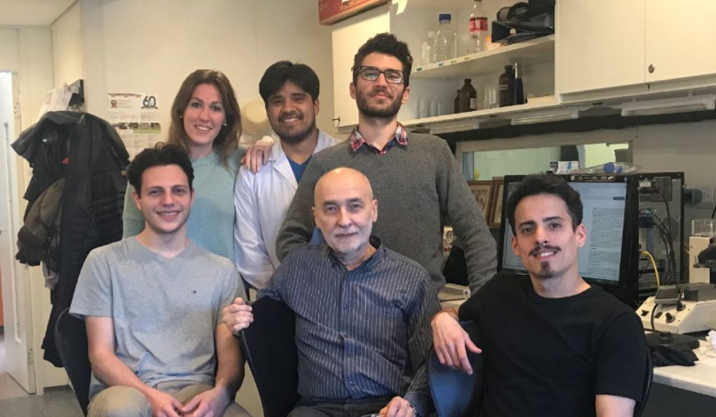
Gender disbalance has been the rule in Argentina, and most conspicuously among professionals, as I witnessed in a male-dominated activity like Medicine. But already during my university days the situation was much better than during my father´s university career. And the disbalance has continued to improve during the last couple of decades, as part of a world phenomenon in the West. If I look at the list of Ph.D. theses and research fellows I have supervised, 75% are women scientists So I would say that I have been lucky that so many female students put their trust in me during such an important professional period of their lives. The subsequent incorporation of many of these students into my research group has had a very positive impact on my scientific career.
What is your favourite type of microscopy and why?
I have had an enchantment with optical microscopy since my childhood. As a graduate student I learnt electron microscopy with my professor of pathology Eduardo Lascano, and then with the person who introduced the electron microscope into Argentina, my thesis supervisor Eduardo De Robertis. My relationship with the electron microscope continud at the Max-Planck-Institute with my friend Peter Zingsheim and with Joachim Frank in the early 1980´s, with whom we performed the first single-molecule imaging of a neurotransmitter receptor. Joachim was awarded the Nobel prize in 2017 for having developed the single-molecule averaging methods. Optical microscopy had a major revolution at the beginning of the 21st. century, when superresolution broke the barrier imposed by the diffraction of light. As I mentioned before, an Alexander von Humboldt award gave me the chance to start a collaboration with Stefan Hell in the late 90´s. Stefan had just returned from Finland and was developing new forms of microscopy. With Stefan we hit another first, imaging the nicotinic acetylcholine receptor with the newly introduced STED superresolution microscopy in 2007. Stefan and his group helped us to build the first superresolution microscope in Argentina in 2008. Stefan Hell was one of the recipients of the Nobel Award in Chemistry in 2014. Optical microscopy has remained my favorite technique, from childhood to the current stage of my scientific career.
What is the most extraordinary thing you have seen by microscopy? An eureka moment for you?
I had a jaw-dropping moment the first time I watched a sample diffract in the electron microscope in La Plata, with my friend Edgardo Macchi, a structural chemist. The diffraction pattern of an organic sample consisted of hundreds if not thousands of intensely brilliant spots that lasted for a few seconds, and then faded under the energy of the bombarding electron beam. It was like a vision of the cosmos projected onto the microscope’s screen. A micrcosmos. Beautiful. Of course the best is yet to come… We are now witnessing resolutions of 1 nm in biological specimens with visible light and conventional lenses! It´s almost unbelievable!
What is an important piece of advice you would give to future Argentinian scientists? and especially those specializing as microscopists?
In the course of my life as a scientist I have witnessed a couple of breakthroughs that were inimaginable 50 years ago. To see biomolecules with an optical microscope, and to be able to track their motion in real time in a living cell, to see a dendritic spine in a neuron dance in a live animal, to image an enzyme with true atomic resolution with cryo-electron microscopy… So my best advice to any scientist, Argentinian or not, is: keep your mind open and enjoy what you do. I have enjoyed what I do in Science all along my career, and find the questions ahead about the functioning of the brain, the origin of life, or the birth of the Universe tremendously exciting. Life started at the micro-scale, so it is easy to predict that micro-scopist will have a niche in the “realm of the small” for years to come…
Where do you see the future of science and microscopy heading over the next decade in Argentina, and how do you hope to be part of this future?
Argentina had a leading role in microscopy in the late 1950´-1960´s, when De Robertis brought the first transmission electron microscope. This led to important developments in the fields of cell biology and structural neurobiology, with the emergence of focal points in Mendoza, Córdoba, La Plata and Tucumán, loan a par with analogous advances elsewhere. For instance, the first Siemens electron microscope in Buenos Aires was up and running within a few years after the one at Oxford University. Today Argentina does not have a single cryo-electron microscope. Superresolution optical microscopy has been implemented in only two or three laboratories, counting ours. If the future is now, Argentina should remember once more Houssay´s motto “Rich countries are so because they invest in scientific-technolgical development, and poor countries remain in that status because they do not invest in these areas”.
Beyond science, what do you think makes Argentina a special place to visit and go to as a scientist?
Argentina is a land with a beautiful and diverse scenery, a rich cultural background, sophisticated cuisine, and above all, interesting people. I have had many foreign visitors, and I cannot think of anyone who has gone back home without numerouspleasant memories of Argentina as a host country.


 (1 votes, average: 1.00 out of 1)