Microscopy preprints – applications in cell biology
Posted by FocalPlane, on 4 November 2022
Here is a curated selection of preprints published recently. In this post, we focus specifically on preprints using microscopy tools in cell biology.
Comparison of interferometric light microscopy with nanoparticle tracking analysis for the study of extracellular vesicles and bacteriophages
Romain Sausset, Zuzana Krupova, Eric Guédon, Sandrine Peron, Alice Grangier, Marie-Agnès Petit, Luisa De Sordi, Marianne De Paepe
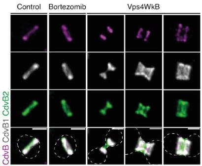
The patterned assembly and stepwise Vps4-mediated disassembly of composite ESCRT-III polymers drives archaeal cell division
Fredrik Hurtig, Thomas C.Q. Burgers, Alice Cezanne, Xiuyun Jiang, Frank Nico Mol, Jovan Traparić, Gabriel Tarrason-Risa, Andre Arashiro Pulschen, Lena Harker-Kirschneck, Anđela Šarić, Rifka Vlijm, Buzz Baum
Particle fusion of Single Molecule Localization Microscopy data reveals dimer structure of Nup96 in Nuclear Pore Complex
Wenxiu Wang, Arjen Jakobi, Yu-Le Wu, Jonas Ries, Sjoerd Stallinga, Bernd Rieger
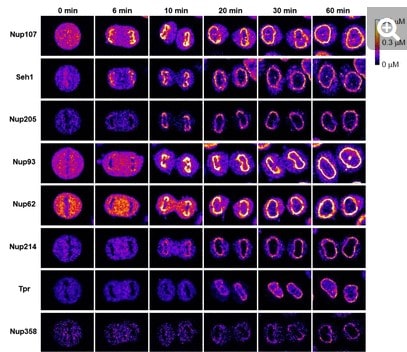
A quantitative map of nuclear pore assembly reveals two distinct mechanisms
Shotaro Otsuka, Jeremy O. B. Tempkin, Wanlu Zhang, Antonio Z. Politi, Arina Rybina, M. Julius Hossain, Moritz Kueblbeck, Andrea Callegari, Birgit Koch, Natalia Rosalia Morero, Andrej Sali, Jan Ellenberg
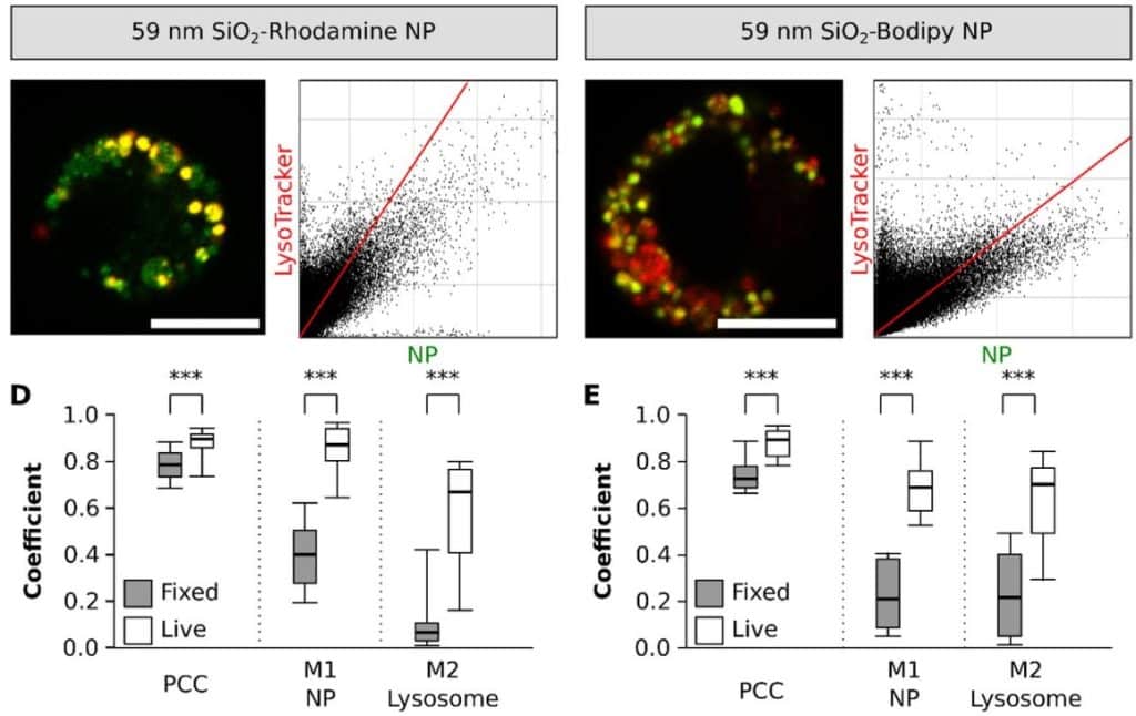
Pitfalls in methods to study colocalization of nanoparticles in mouse macrophage lysosomes
Aura Maria Moreno-Echeverri, Eva Susnik, Dimitri Vanhecke, Patricia Taladriz-Blanco, Sandor Balog, Alke Petri-Fink, Barbara Rothen-Rutishauser
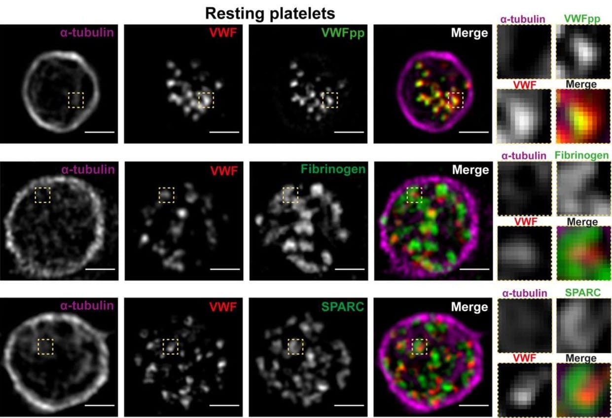
Quantitative super-resolution imaging of platelet degranulation reveals differential release of VWF and VWF propeptide from alpha-granules
Maurice Swinkels, Sophie Hordijk, Petra E. Bürgisser, Johan A. Slotman, Tom Carter, Frank W.G. Leebeek, A.J. Gerard Jansen, Jan Voorberg, Ruben Bierings
Single-molecule imaging of cytoplasmic dynein in cellulo reveals the mechanism of motor activation and cargo movement
Nireekshit Addanki Tirumala, Gregory Redpath, Sarah Viktoria Skerhut, Pritha Dolai, Natasha Kapoor-Kaushik, Nicholas Ariotti, K Vijay Kumar, Vaishnavi Ananthanarayanan
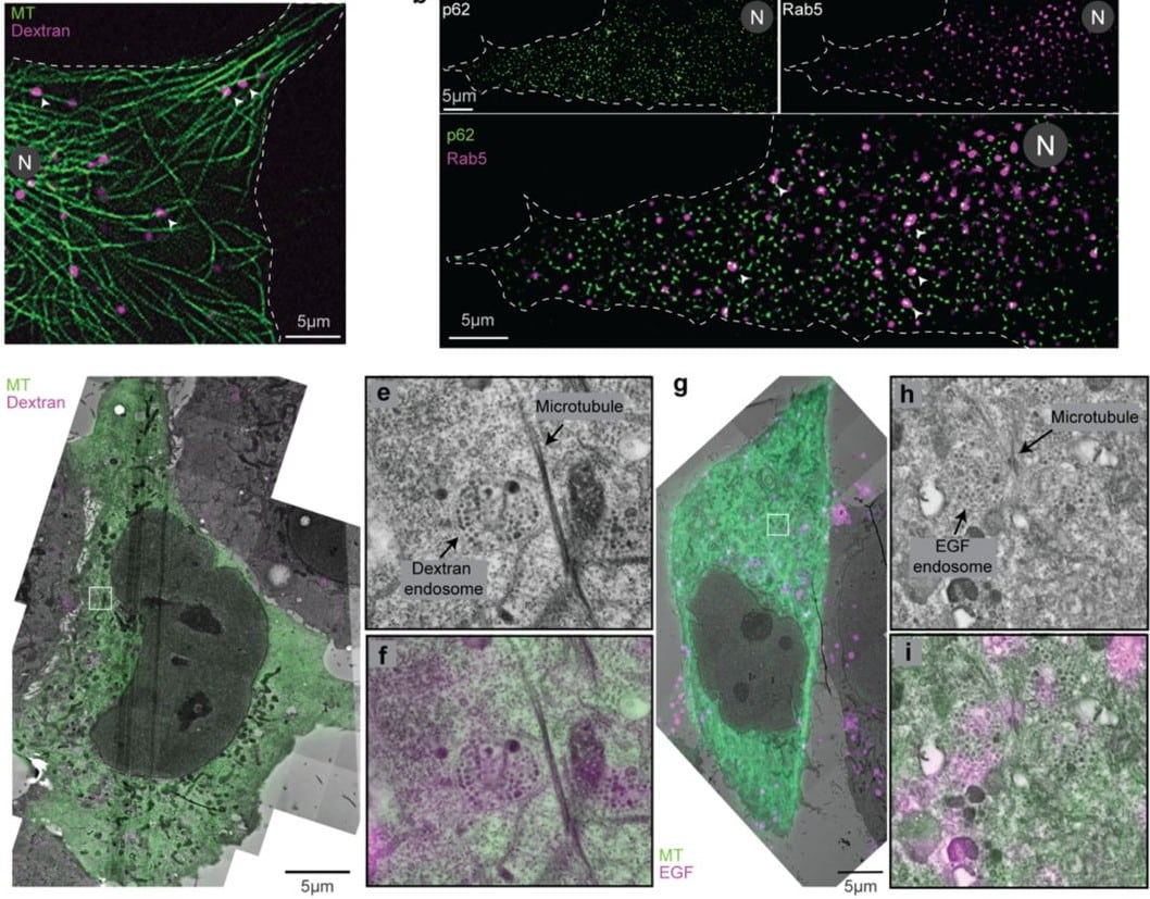
Unraveling the intricate microtubule inner protein networks that reinforce mammalian sperm flagella
Miguel Ricardo Leung, Marc C. Roelofs, Riccardo Zenezini Chiozzi, Johannes F. Hevler, Albert J. R. Heck, Tzviya Zeev-Ben-Mordehai
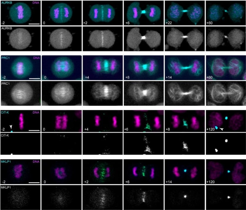
Midbody proteins display distinct temporal dynamics during cytokinesis
Ella F.J. Halcrow, Riccardo Mazza, Anna Diversi, Anton Enright, Pier Paolo D’Avino
A balance between actin and Eps8/IRSp53 utilization in branched versus linear actin networks determines tunneling nanotube formation
J. Michael Henderson, Nina Ljubojevic, Thibault Chaze, Daryl Castaneda, Aude Battistella, Quentin Giai Gianetto, Mariette Matondo, Stéphanie Descroix, Patricia Bassereau, Chiara Zurzolo
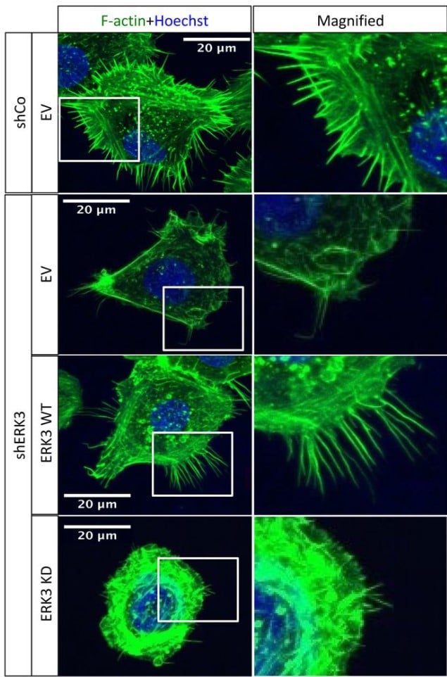
ERK3/MAPK6 dictates Cdc42/Rac1 activity and ARP2/3-dependent actin polymerization
Katarzyna Bogucka-Janczi, Gregory Harms, Mary May-Coissieux, Mohamad Bentires-Alj, Bernd Thiede, Krishnaraj Rajalingam
Multi-modal mass spectrometry imaging reveals single-cell metabolic states in mammalian liver
Hua Tian, Presha Rajbhandari, Jay Tarolli, Aubrianna M. Decker, Taruna V. Neelakantan, Tina Angerer, Fereshteh Zandkarimi, Jacob Daniels, Helen Remotti, Gilles Frache, Nicholas Winograd, Brent R. Stockwell
Heterogeneous and Surface-Catalyzed Amyloid Aggregation Monitored by Spatially Resolved Fluorescence and Single Molecule Microscopy
Xin Zhou, Anders Wilgaard Sinkjær, Min Zhang, Henrik Dahl Pinholt, Hanne Mørck Nielsen, Nikos S. Hatzakis, Marco van de Weert, Vito Foderà
Multifocal two-photon excitation fluorescence microscopy reveals hop diffusion of H-Ras membrane anchors in epidermal cells of zebrafish embryos
Radoslaw J. Gora, Redmar C. Vlieg, Sven Jonkers, John van Noort, Marcel J.M. Schaaf
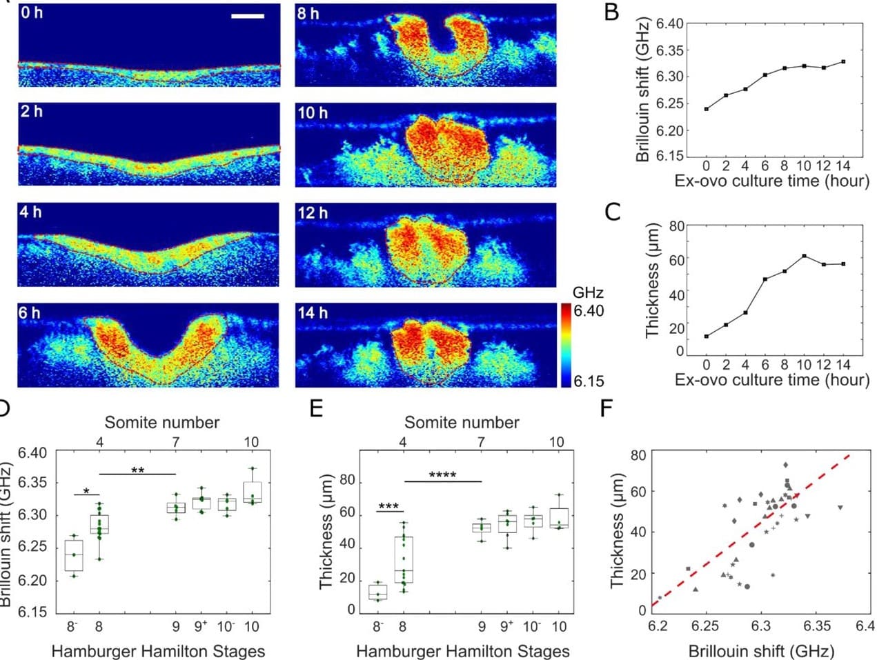
Time-lapse mechanical imaging of neural tube closure in live embryo using Brillouin microscopy
Chenchen Handler, Giuliano Scarcelli, Jitao Zhang
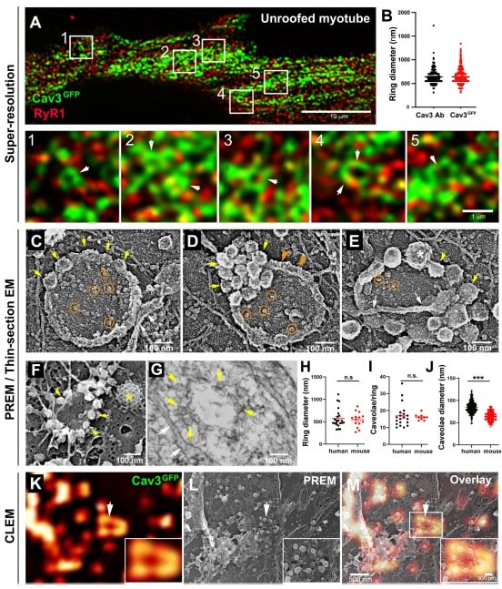
Caveolae and Bin1 form ring-shaped platforms for T-tubule initiation
Eline Lemerle, Jeanne Lainé, Gilles Moulay, Anne Bigot, Clémence Labasse, Angéline Madelaine, Alexis Canette, Perrine Aubin, Jean-Michel Vallat, Norma B Romero, Marc Bitoun, Vincent Mouly, Marty Isabelle, Bruno Cadot, Laura Picas, Stéphane Vassilopoulos
 (No Ratings Yet)
Get involved
Create an account or log in to post your story on FocalPlane.
More posts like this

Filter by
- NewsApply
- DiscussionsApply
- How toApply
- ToolsApply
- Case studiesApply
- InterviewsApply
- JobsApply
- EducationApply
- Blog seriesApply
- WAMBIAN: West Africa.. in FocusApply
- Volume EMApply
- Latin American Micro..scopistsApply
- Bio-image Analysis w..ith NapariApply
- Imaging with…Apply
- Towards Global Acces..sApply
- Latin America Bioima..gingApply
- From Zero to Qupath ..HeroApply
- Asian Microscopists ..and Cell BiologistsApply
- AIC at HHMI JaneliaApply
- Deep Learning for Bi..o-image analysisApply
- GloBIAS – updates fr..om the communityApply
- Highlights from Euro..-BioImagingApply
- LSFM seriesApply
- DIY MicroscopyApply
- View all