Image checklists v1.0 from QUAREP-LiMi
Posted by Helen Zenner, on 16 March 2023
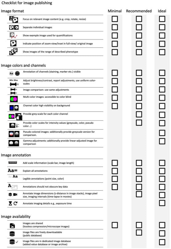
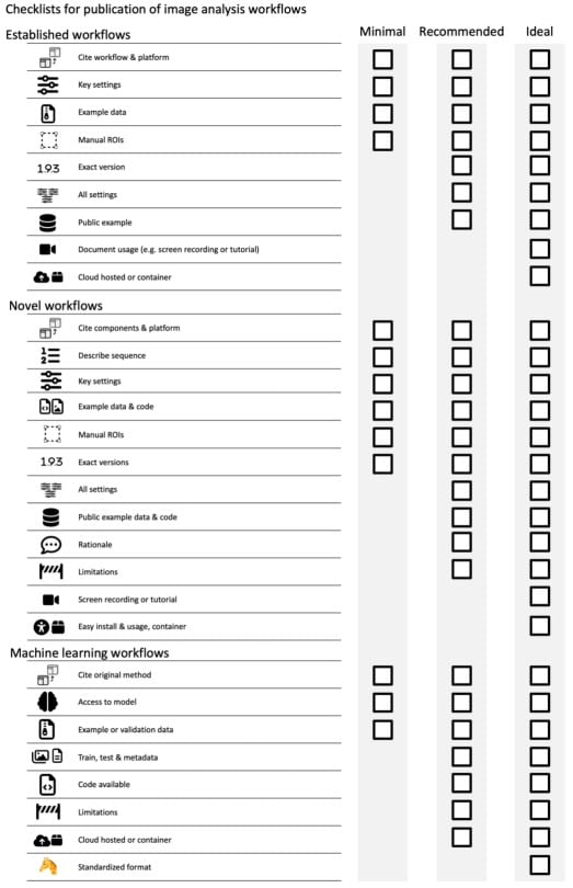
QUAREP-LiMi (Quality Assessment and Reproducibility for Instruments and Images in Light Microscopy) is a community group of light microscopists, science communicators, business representatives, vendors and application specialists aiming to improve both quality assessment and quality control in microscopy. QUAREP-LiMi is organised into (currently) 13 working groups, focusing on areas such as illumination power, lateral and axial resolution and phototoxicity. Recently, the ‘Image analysis and visualization’ working group led by Helena Jambor and Christopher Schmied published guidelines to ensure quality, consistency and reproducibility of microscopy data in publications. The guidelines themselves come in the form of two handy checklists, one on image publishing and the second on documenting image analysis workflows. These checklists are designed for everyone, from users that are new to microscopy to experts in imaging facilities, as well as recommendations for publishers.
I caught up with Helena Jambor and Alex Payne-Dwyer, two of the 54 authors, with some questions about the checklists and how they see the guidelines evolving in the future.
Why is having these guidelines so important?
HJ: “Microscope images have evolved from photographs to digital, quantitative data. The undergraduate curricula and publishing guidelines however have not quite been updated to include ‘how-to’ for digital image presentation in papers. It is therefore high time to set standards and our checklists are a first step.
Was it difficult to come to a consensus?
HJ: “Oh yes! For more than one year we discussed point by point all aspects in a ton of detail. But we often quickly agreed on the checklist items but needed time to discuss the details. It was much less difficult to agree on our goals, and more challenging to first agree on terminologies and semantics!”
Why do you have three levels, minimal; recommended and ideal – why not just go with ideal?
HJ: “We wanted to find a way to introduce really easy-to-achieve goals that could accommodate as many researchers and publishers as possible. E.g., submitting to image archives is laborious and needs resources and know-how that not available everywhere. These are the minimal guidelines. However, we also wanted to make it clear that we believe more should be put into place, these are the recommended guidelines. And last, the sky is the limit! In a few years reproducible science hopefully will be mainstream, so maybe then our dreams and ideal guidelines will become feasible!
You mention that the guidelines will evolve over time, is there anything on the horizon that we should be looking for?
HJ: “We are a diverse group and have quite a number of interesting ideas for the future. A couple of us are planning to continue with educational materials within the QUAREP-LiMi scope, e.g., video tutorials etc. My co-chair Christopher Schmied is dreaming of a living document that expands and evolves over time as methods also continue to be developed, especially in the image analysis arena. At the end for most of us this is voluntary work, so we also have to see what is realistic.”
A P-D: “We often hear about how AI will be disruptive to scientific reproducibility and innovation. This is already happening, not only with the rise of generative imaging and description models (notoriously in AI art), but also with more impressive tools for interpolation of quantitative images, especially to combat phototoxicity and noise. The current checklist begins to address the complex impact of AI on post-processing and visualisation. However, we don’t often recognise that AI approaches will also impact the acquisition of images, known as ‘smart’ microscopy. Not every image needs to be taken – just those which provide information on the research question. Another field of active development is the accessibility and interactivity of published microscopy data beyond 2D print, for example with augmented and virtual visualisation of high-dimensional images at terabyte scale.”
Below are the ‘titbits from the text’ that I thought were important/I didn’t know, followed by my take-home messages
- Rotating images through angles other than multiples of 90°alters intensity values through interpolation
- ALWAYS make changes on a duplicate of the original data
- Grayscale colour schemes are uniformly perceived
- Save your data using lossless compression
- Making your data ‘available on request’ is an inefficient method of sharing data
Take-home messages
- it is essential to know what you are doing to your image, whether this is when preparing a figure or when going through processing steps during bioimage analysis
- make sure that any colour schemes and annotations are clear and accessible for all
- record what you are doing – either the metadata linked to the images or during image processing so someone can reproduce your imaging or analysis
- use image repositories (such as OMERO, BioImage Archive, EMPIAR) and code repositories (such as github), with caveats around the size requirements for some imaging modalities
 (2 votes, average: 1.00 out of 1)
Get involved
Create an account or log in to post your story on FocalPlane.
More posts like this


Filter by
- NewsApply
- DiscussionsApply
- How toApply
- ToolsApply
- Case studiesApply
- InterviewsApply
- JobsApply
- EducationApply
- Blog seriesApply
- Latin America Bioima..gingApply
- From Zero to Qupath ..HeroApply
- Asian Microscopists ..and Cell BiologistsApply
- AIC at HHMI JaneliaApply
- Deep Learning for Bi..o-image analysisApply
- GloBIAS – updates fr..om the communityApply
- WAMBIAN: West Africa.. in FocusApply
- Volume EMApply
- Latin American Micro..scopistsApply
- Bio-image Analysis w..ith NapariApply
- Imaging with…Apply
- Towards Global Acces..sApply
- Highlights from Euro..-BioImagingApply
- LSFM seriesApply
- DIY MicroscopyApply
- View all