New Features & Reviews Editor at Journal of Cell Science
Posted by Amelia Glazier, on 4 April 2023
Dear FocalPlane Community,
My name is Amelia Glazier and I have recently joined the Company of Biologists as Features & Reviews Editor for Journal of Cell Science. During my PhD, I developed a strong interest in ethical scholarly publishing and scientific communication. Now I am transitioning from bench research to the scientific publishing field, and I am very honoured and excited to begin helping researchers communicate their cutting-edge contributions to cell biology. I will be working with the excellent editors at Journal of Cell Science to publish Reviews, Cell Science at a Glance posters, Research Highlights, interviews with authors and up-and-coming cell biologists, and more. As part of my role, I will be joining the FocalPlane team.
My research background focused on protein homeostasis mechanisms in the context of cardiac and skeletal muscle disease. My first experiences in the lab as an undergraduate exploring effects of post-translational modifications of sarcomere proteins, with Dr. Lori Walker at the University of Colorado Denver and Dr. Brandon Biesiadecki at the Ohio State University, introduced me to the amazing complexity of the sarcomere. I have been hooked on striated muscle cell biology ever since. I completed my PhD in Molecular and Integrative Physiology at the University of Michigan supervised by Drs Sharlene Day and Daniel Michele, studying protein degradation pathways and sarcomere-chaperone interactions in hypertrophic cardiomyopathy associated with mutations in cardiac myosin binding protein C. To further broaden my knowledge of protein quality control mechanisms and experimental models, I recently completed postdoctoral work at the University of Ulm in Dr. Steffen Just’s lab, investigating roles of endolysosomal and autophagic pathways in cardiomyopathy and cardiac aging using zebrafish models.

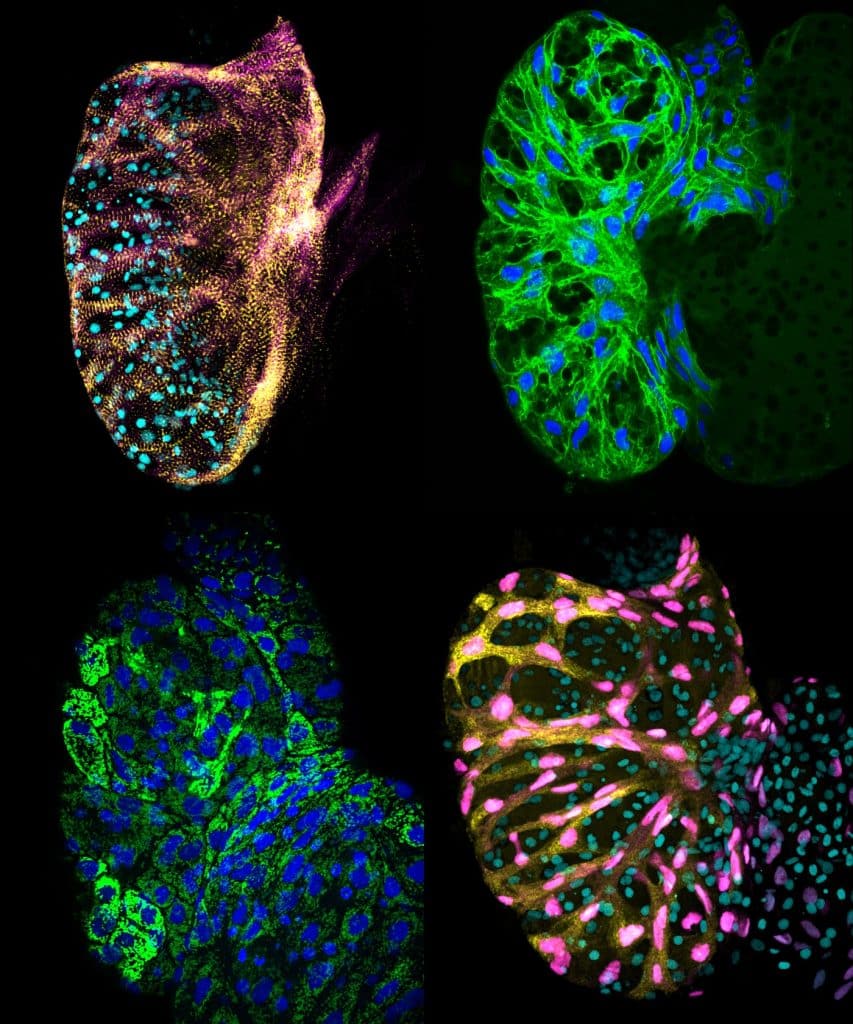
Clockwise from top left: Z-disk (ttn.2:actn2-EGFP;myl7:mCherry-CAAX); sarcolemma (unc45bmin:GFP-CAAX); mitochondria (ef1a:MLS-EGFP); myonuclei and sarcoplasm (myl7:mCherry-NLS;vmhc:EGFP).
Understanding the elaborate maintenance of the sarcomere required for healthy muscle function remains a challenging task. Ever since the publication of the first electron micrographs of isolated myofibrils inspired and supported the ground-breaking sliding filament hypothesis of muscle contraction, microscopy techniques have helped advance our conceptualization of the sarcomere from a largely static mechanical lattice to a dynamic and sensitively regulated machine perpetually under construction. More recently, click chemistry has allowed visualization of spatiotemporal sarcomere protein synthesis and degradation (Lewis et al 2018, Scarborough et al 2021), FRAP lets us observe sarcomere protein turnover in real time (Leber et al 2016, Rudolph et al 2019), and dSTORM can paint a picture of sarcomere components in stunning resolution (Szikora et al 2019, Previs et al 2015).
And while I of course appreciate the scientific utility of microscopy in muscle cell biology, I have also always simply found cardiomyocytes and the sarcomere beautiful to image! During my postdoc, I often used confocal microscopy to visualize different structures in the embryonic hearts of transgenic zebrafish.

I am very much looking forward to working with the FocalPlane community and with the greater cell biology field! Please do not hesitate to contact me at amelia.glazier@biologists.com for inquiries regarding front section content for Journal of Cell Science.
Amelia Glazier, PhD
Features & Reviews Editor, Journal of Cell Science
The Company of Biologists, Cambridge, UK


 (2 votes, average: 1.00 out of 1)