Imaging with the Liverpool Centre for Cell Imaging (CCI)
Posted by FocalPlane, on 7 September 2023
In the first of our series of ‘Imaging with…’ blog posts, we meet the staff at the Liverpool CCI
Staff role call:
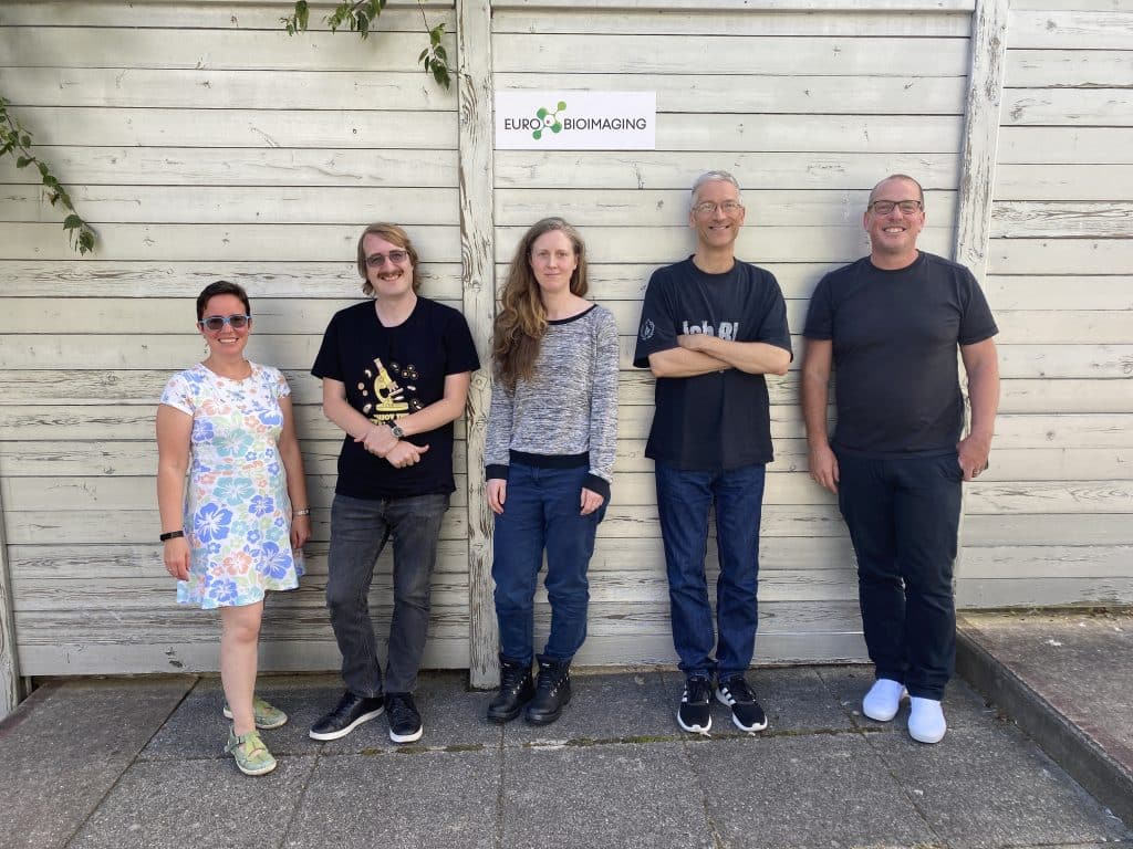
Tobias Zech, Academic lead
Most likely to be found behind his desk writing applications and making exciting (in his own words) plans for the future of the CCI and imaging in general at the University of Liverpool.
Marco Marcello, Facility Manager
Expertise: developing/working on microscopes since 2000
Most likely to be found with his eyes glued to oculars or computer screens.
Thomas Waring, Imaging Technician
Expertise in experimental design and light microscopy
Most likely to be found in a darkened room, vibing to some music while taking pretty pictures on the microscopes.
Lucy Isherwood, Imaging Technician
Expertise in light and electron microscopy
Most likely to be found speedily carrying samples between the CCI and EM facility.
Marie Held, Image Analyst
Expertise in generating numbers from images with a variety of software packages
Most likely to be found at her desk analysing images or numbers whilst nursing a cup of green tea.
Microscope role call:
Zeiss Elyra Lattice SIM & PALM/STORM, Zeiss/Bruker LSM880-BioAFM; Zeiss LSM880-Multiphoton; Zeiss LSM780-FLIM ; Zeiss LSM900-Airyscan2; Zeiss LSM800-Airyscan; Leica SP5; Light sheet (Zeiss Z1); 3i Marianas spinning disk confocal-TIRF; Andor spinning disk confocal Dragonfly-SRRF; 2 x Incucyte S3 medium throughput live cell imaging; Zeiss CellDiscoverer 7-Airyscan2 high throughput live cell imaging; Widefield (Zeiss ×2).
Who can access the CCI?
We are open to everybody. We train users in independent microscope use and image analysis or perform full projects as service work, depending on user inclination, available time and funds. We have recently been selected, along with six leading institutions, as member of the UK Node of Euro-Bioimaging that makes it very easy for international users to access and book our resources (https://www.eurobioimaging.eu/about-us/how-to-access). The CCI works in close collaboration with the Biomedical Electron Microscopy unit and the Centre for Pre-clinical Imaging to enable the best imaging solutions across scales.
The following section has been answered by Marie Held (Image Analyst) and represents her personal opinion
Pet peeve in microscope users
Leaving the microscope with the stage not centred and not with the lowest magnification objective in the light path.
Favourite microscope
Lightsheet Z.1 but closely followed by the Elyra 7 Lattice SIM.
Favourite thing to image
3D cultures grown with or without scaffolds.
Best bit of advice (that you give or have been given)
Think about your intended analysis before you start imaging, even better, whilst planning the experiment.
If money was no object, I would buy…
A PC with the most powerful graphics card fashioned with the best performance, high resolution screen that I can find situated on the most suitable hybrid (standing/siting) desk, desk chair and PC periphery for my short physique.
Can you give us an example of a recent paper with the assistance of CCI: “As we cannot make up our minds, we are offering two exemplary publications.”
- Manning, Declan, Richard Barrett-Jolley, Richard L. Evans, and Caroline Dart. ‘TRPC1 Channel Clustering during Store-Operated Ca2+ Entry in Keratinocytes’. Frontiers in Physiology 14 (2023). https://www.frontiersin.org/articles/10.3389/fphys.2023.1141006.
- Hannah Elcocks, Ailbhe J Brazel, Katy R McCarron, Manuel Kaulich, Koraljka Husnjak, Heather Mortiboys, Michael J Clague, and Sylvie Urbé. ‘FBXL4 Ubiquitin Ligase Deficiency Promotes Mitophagy by Elevating NIX Levels’. The EMBO Journal (2023) 42:e112799. https://doi.org/10.15252/embj.2022112799.
How should users acknowledge the facility and why is it important?
Acknowledgement in publications and presentations of CCI performed work is vital to secure support and funding necessary to maintain this valuable research resource. For publications that include work performed in the facility, please use the acknowledgement statement “We acknowledge the Liverpool Centre for Cell Imaging (CCI) for provision of imaging equipment and technical assistance.” and include required grant numbers as listed on the facility webpage. Please also consider staff for a named acknowledgement mention or even co-authorship if they played a key role in the study. If in doubt, consult the community-developed RMS Imaging Facility Guidelines for Acknowledgement.
 (3 votes, average: 1.00 out of 1)
Get involved
Create an account or log in to post your story on FocalPlane.
More posts like this

Filter by
- NewsApply
- DiscussionsApply
- How toApply
- ToolsApply
- Case studiesApply
- InterviewsApply
- JobsApply
- EducationApply
- Blog seriesApply
- GloBIAS – updates fr..om the communityApply
- WAMBIAN: West Africa.. in FocusApply
- Volume EMApply
- Latin American Micro..scopistsApply
- Bio-image Analysis w..ith NapariApply
- Imaging with…Apply
- Towards Global Acces..sApply
- Latin America Bioima..gingApply
- From Zero to Qupath ..HeroApply
- Asian Microscopists ..and Cell BiologistsApply
- AIC at HHMI JaneliaApply
- Deep Learning for Bi..o-image analysisApply
- Highlights from Euro..-BioImagingApply
- LSFM seriesApply
- DIY MicroscopyApply
- View all