Imaging with the Instituto Gulbenkian de Ciência’s Electron Microscopy Facility
Posted by FocalPlane, on 14 December 2023
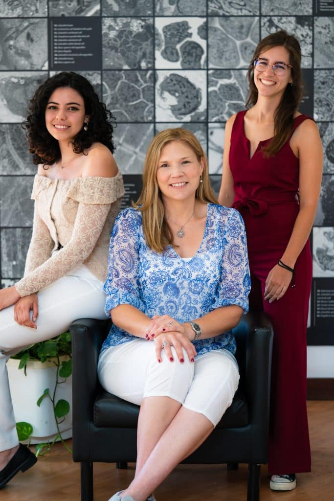
In our ‘Imaging with…’ blog post, we meet the staff of the Electron Microscopy (EM) facility at the Instituto Gulbenkian de Ciência, Lisbon, Portugal.

Staff role call:
Erin Tranfield, Facility Head
Expertise: Fire fighting
Most likely to be found lost in a Zoom meeting
Ana Laura Sousa, Senior Technician
Expertise: “Everything, everywhere, all at once” but nothing at all, since in science we are always learning.
Most likely to be found singing in the facility
Beatriz Tomaz, Junior Technician
Expertise: A ridiculous amount of Negative Staining
Most likely to be found trying to do everything at once
Microscope role call:
One and a half microscopes (TEM FEI Tecnai G2 Spirit BioTWIN and old guy TEM Hitachi H-7650).
Who can access the facility?
We welcome anyone who wants to exchange ideas, brainstorm about electron microscopy and learn about the powerful tool that is electron microscopy – whether they are from within our institute or from other institutes and companies.
Pet peeve (something that users do that is annoying)
We wouldn’t say annoying, but we are not too fond of people dropping by without booking in advance or put in requests at 5pm as we are about to head home!
Favourite microscope
This answer can be controversial, while the Hitachi is certainly the old favourite, there’s some features only the FEI can provide.
Favourite thing to image
We get such a wide range of samples it’s hard to say! Bee gut can be fun, and viruses are also super interesting.
Best bit of advice (that you give or have been given)
No feedback is good feedback!!! – by Erin Tranfield herself
If money was no object, we would buy…
vSEM, vSEM, vSEM, did we mention a vSEM? Dear Santa, we would really like a volume SEM please and thank-you! If money was really not an issue, we would also ask Santa for another 120kV TEM, and a new 200kV TEM, and of course additions to the team to operate these new toys!
Can you give us some examples of recent papers that were published with your assistance?
- The 3D architecture and molecular foundations of de novo centriole assembly via bicentrioles (2021) DOI: 10.1016/j.cub.2021.07.063
- ATG9A regulates the dissociating of recycling endosomes from microtubules to form liquid influenza A virus inclusions (2023) DOI: 10.1371/journal.pbio.3002290
How should users acknowledge the facility and why is it important?
Our institute had the policy that all users of the facilities should acknowledge the support of the core facilities. Despite this request, we are always thankful of users that take the time to acknowledge our contribution to their research in publications and presentations. To help we offer a template our users can use, stating the name of the facility, of our institute and of the technician who worked on the project.
A simple acknowledgement can really show to our institute and the broad scientific community how much impact Electron Microscopy can have in research and particularly how our facility plays a role in this. Overall, this also showcases the satisfaction from our users and potentially brings in more funding and community recognition for the facility.
All this helps the facility gain a good reputation and of course, at a personal level, for each of us it’s important and meaningful to have our expertise and invested time rewarded and recognised.


 (5 votes, average: 1.00 out of 1)