Targeted vCLEM of heterogeneous samples
Posted by Mario Elias Ortega Sandoval, on 15 January 2024
by Karel Mocaer* and Paolo Ronchi#
*Schwab Team and #EMCF, EMBL Heidelberg, Germany
Challenge
Studying cells or events within their biological context (i.e. in tissues, ecosystem etc…) is highly relevant. However, volume EM techniques are limited in the volume that can be acquired. Therefore, targeting approaches are needed to characterize specific regions of interest. This is particularly important when investigating rare events in a heterogeneous sample. To overcome this bottleneck we have developed a targeting approach based on the fluorescence of exogenously tagged proteins1, organic dyes2, or autofluorescence3.
Technique: LM guided FIB-SEM imaging
Focused ion beam – scanning electron microscopy (FIB-SEM) allows the acquisition of vEM data by combining iterative block surface imaging and ablation of thin layers of resin with an ion beam, thus providing 3D subcellular information. In order to target specific fluorescent events in an heterogeneous sample, we have developed a workflow that relies on the preservation of fluorescence when processing the sample for EM. We can then acquire a fluorescent 3D map of the resin block using a confocal microscope (e.g. Fig.1, reproduced from 3) and use it to efficiently target the vEM acquisition. As light microscopy and EM are performed on the same block post embedding, we can overlay the 2 datasets with high precision, thus associating molecular information with morphology (e.g. in Fig. 1, chlorophyll fluorescence in magenta overlaps with the chloroplast in a dinoflagellate. Reproduced from 3)
Research
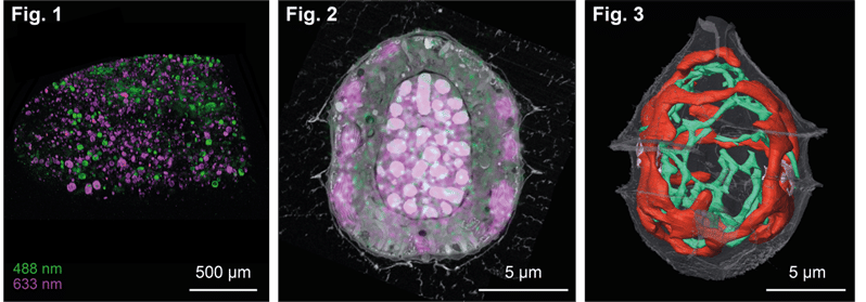
Using this method we could acquire a specific cell of interest from an heterogeneous marine environmental sample (Fig. 1) and perform a morphometric analysis of various organelles (Fig. 2, chloroplast in red and mitochondrion in green). The same workflow has also been applied to target fluorescent cells in Drosophila tissues1 and organoids1,2.
Impact
•FIB-SEM allows for high resolution subcellular ultrastructural analysis of cells. From this we can extract quantitative volume data.
•Preservation of fluorescence or autofluorescence allows for precise targeting of cells of interest in their environment.
References
1Ronchi et al, JCB, 2021;
2D’imprima et al, Dev Cell, 2023, 3Mocaer et al, JCS, 2023
 (No Ratings Yet)
Get involved
Create an account or log in to post your story on FocalPlane.
More posts like this


Filter by
- NewsApply
- DiscussionsApply
- How toApply
- ToolsApply
- Case studiesApply
- InterviewsApply
- JobsApply
- EducationApply
- Blog seriesApply
- Volume EMApply
- Latin American Micro..scopistsApply
- Bio-image Analysis w..ith NapariApply
- Imaging with…Apply
- Towards Global Acces..sApply
- Latin America Bioima..gingApply
- From Zero to Qupath ..HeroApply
- Asian Microscopists ..and Cell BiologistsApply
- AIC at HHMI JaneliaApply
- Deep Learning for Bi..o-image analysisApply
- GloBIAS – updates fr..om the communityApply
- WAMBIAN: West Africa.. in FocusApply
- Highlights from Euro..-BioImagingApply
- LSFM seriesApply
- DIY MicroscopyApply
- View all