Imaging with The University Imaging Centers, a Nikon Center of Excellence, at the University of Minnesota – Twin Cities
Posted by FocalPlane, on 25 January 2024
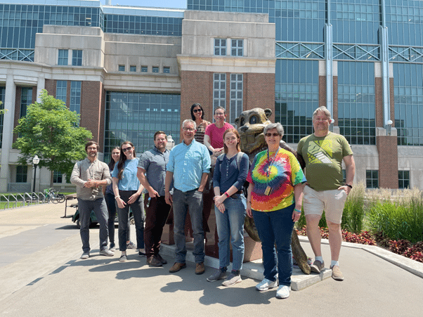
In our ‘Imaging with…’ blog post, we meet the staff of The University Imaging Centers, a Nikon Center of Excellence, at the University of Minnesota – Twin Cities.

Staff role call
Dr Mark Sanders, Program Director
Expertise: Biological microscopy and imaging, core facility management
Most likely to be found on Zoom troubleshooting or in meetings, or fishing.
Grant M. Barthel, Assistant Director and Spatial Biology Lead
Expertise: Spatial transcriptomics and proteomics, light microscopy
Most likely to be found troubleshooting.
Dr Guillermo Marques, Scientific Director
Expertise: confocal, multiphoton, widefield, and TIRF microscopy and structured illumination microscopy
Most likely to be found in front of a microscope helping a client.
Alexander Cramer, Operations Manager
Expertise: Administration, Communications, Reporting, Customer Support
Most likely to be found working with data in Tableau.
Gregg Amundson, IT Support
Expertise: Computer and Network configuration
Most likely to be found debugging a script.
Dr Mary Brown, Imaging Specialist and Lead Image Data Analyst
Expertise: Confocal microscopy and image processing & analysis
Most likely to be found in front of a computer or workstation, sporting either a Michigan or Minnesota football shirt.
Dr Gail Celio, Electron Microscopy Section Lead
Expertise: SEM, TEM, specimen preparation, ultramicrotomy
Most likely to be found sectioning or putting cool decals on scientific instruments.
Erin Hudson, Spatialomics and Imaging Specialist
Expertise: Spatial transcriptomics and proteomics
Most likely to be found at the lab bench.
Dr Jason Mitchell, Imaging Specialist
Expertise: 2-photon, light microscopy, small animal (intravital, ultrasound, PET, uCT)
Most likely to be found in the hallway walking between facilities.
Anna Peyla, Spatialomics and Imaging Specialist
Expertise: RNAscope processing and imaging
Most likely to be found training new users on the Nikon AX R.
Laurel Schuck, Tissue Clearing and Imaging Specialist
Expertise: Clearing of whole tissue volumes, light sheet imaging of cleared tissue, confocal microscopy
Most likely to be found at the lab bench.
Steve Schnell, Small Animal Imaging and Imaging Specialist
Expertise: Small animal imaging (MRI, ultrasound, chemiluminescence), confocal microscopy, image processing and data analysis.
Most likely to be found helping our clients and learning something new.
Patrick Willey, Tissue Clearing and Imaging Specialist
Expertise: Imaging and processing large samples, troubleshooting strange microscope malfunctions, and teaching users how to acquire their best data
Most likely to be found training a new user on a ‘scope or imaging a cleared mouse brain
Microscope role call:
Jackson Hall: Nikon A1R HD Multiphoton Microscope, Nikon A1Rsi HD Confocal with SIM Super-Resolution, Olympus FluoView FV1000 BX2 Upright Confocal, Olympus FluoView FV1000 IX2 Inverted Confocal with FLIM Detector, Zeiss Cell Observer Spinning Disk Confocal, 3i Cleared Tissue Light-Sheet, Nikon AZ100M Fluorescence Macroscope, Panoptiq Digital Slide Imaging System, Zeiss Widefield and TIRF Microscope, IncuCyte SX5 Live-Cell Imaging System, IVIS Spectrum In Vivo Imaging System, Vevo 2100 Ultra High-Frequency Ultrasound, Aspect Imaging M5 Compact MRI, Huron TissueScope Slide-Scanning System, Typhoon FLA 9500, and Amersham Imager 600 UV.
Cancer and Cardiovascular Research Building: Nikon AX R, Nikon C2+ Upright Spectral Confocal and Widefield Imaging System, Caliber ID RS-G4 Ribbon-Scanning Confocal, Leica SP5 Upright Confocal Microscope, Leica LMD6500 Laser Dissection Microscope, Leica MZ FL III Fluorescence Stereomicroscope, Nikon Ti2 Microscope, Nikon Ti-E Inverted Deconvolution Microscope, IVIS Spectrum In Vivo Imaging System, Sofie G8 microPET/CT, and Vevo F2.
Spatial Biology Tools: Ionpath MIBIscope, Nanostring GeoMx, Nanostring CosMx, and 10X Genomics Cytassit.
Snyder Hall: Nikon AX R Confocal Microscope System, Nikon A1si Confocal and Widefield Microscope, Nikon AZ100 C1si Spectral Confocal Microscope, Nikon 90i Widefield microscope, JEOL JEM-1400Plus Transmission Electron Microscope (TEM), Hitachi S-3500N Variable Pressure Scanning Electron Microscope (SEM), and Maestro In-Vivo Imaging System.
Who can access the facility?
The University Imaging Centers (UIC) services the entire University of Minnesota plus external clientele beyond the university. We strive to educate our clientele in rigorous, reproducible microscopy and image analysis. UIC staff (13 strong) provide microscope training and image analysis training and services. We also educate researchers with workshops on confocal microscopy and image analysis. In addition to extensive microscopy services, we provide a tissue clearing service along with imaging on a Cleared Tissue Light-Sheet microscope (3i), a ribbon-scanning confocal (Caliber), and a Nikon A1R MP confocal/multiphoton system. We also offer brightfield and widefield fluorescent slide-scanning services. We have contributed to over 60 publications and numerous grant proposals over the past two years. The UIC is one of sixteen Nikon Centers of Excellence in the Americas and one of the largest core facilities in the Midwest. More information can be found at https://med.umn.edu/uic.
Pet peeve (something that users do that is annoying): Unrealistic deadlines/expectations; “Is it on?”; not using proper positive & negative controls
Favourite microscope: Anything Nikon; 3i Cleared Tissue Light-Sheet Microscope; Leica Stellaris 8; Hitachi S-3500N SEM
Favourite thing to image: Anything with strong quantifiable signals in multiple channels; anything without chlorophyll; mammalian cytoskeleton
Best bit of advice (that you give or have been given): Good images start with good sample prep; don’t assume clients fully know what they’re doing or what they want, but always be polite about it; it’s going to take longer than you think
If money were no object, we would buy… More staff; espresso machine; couch; supercomputer & server for big data.
Can you give us some examples of recent papers that were published with your assistance?
Nicholas W. Kreofsky, Punarbasu Roy, Mary E. Brown, Ulises Perez, Ryan E. Leighton, Renee R. Frontera, and Theresa M. Reineke. Cinchona Alkaloid Polymers Demonstrate Highly Efficient Gene Delivery Dependent on Stereochemistry, Methoxy Substitution, and Length. Biomacromolecules 2024; 25 (1): https://doi.org/10.1021/acs.biomac.3c01099
KY Song, YH Han, H Roerich, ME Brown, C Torres-Cabala, and A Giubellino. MET Receptor Tyrosine Kinase Inhibition Reduces Interferon-Gamma (IFN-γ)-Stimulated PD-L1 Expression through the STAT3 Pathway in Melanoma Cells. Cancers 2023; 15 (13): https://doi.org/10.3390/cancers15133408
Hongming Su, Hong Guo, Xiaoxue Qiu, Te-Yueh Lin, Chao Qin, Gail Celio, Peter Yong, Mark Sanders, Xianlin Han, David A. Bernlohr & Xiaoli Chen. Lipocalin 2 regulates mitochondrial phospholipidome remodeling, dynamics, and function in brown adipose tissue in male mice Lipocalin 2 regulates mitochondrial phospholipidome remodeling, dynamics, and function in brown adipose tissue in male mice. Nat Commun 2023; 14, 6729. https://doi.org/10.1038/s41467-023-42473-2
Nicholas Greatens, Nick Klejeski, Les J. Szabo, Yue Jin, and Pablo D. Olivera. Puccinia coronata var. coronata, a Crown Rust Pathogen of Two Highly Invasive Species, Is Detected Across the Midwest and Northeastern United States. Plant Disease 2023; 107(10), 2009. https://apsjournals.apsnet.org/doi/10.1094/PDIS-07-22-1711-RE
How should users acknowledge the facility and why is it important?
For purposes of funding and justification, the University Imaging Centers should be acknowledged in research publications for which imaging or image analysis was performed at the UIC. Such acknowledgment may be written as “This work was supported by the resources and staff at the University of Minnesota University Imaging Centers, SCR_020997.” Staff may assist with text descriptions of instrumentation and imaging or analysis methods in publications. When staff provide additional services, such as developing new sample preparation protocols, design or redesign of experimental conditions, constructive data analysis, or developing image analysis tools such as macros or Nikon Elements General Analysis routines, then the assisting staff should be included in the author list of the publication. Detailed guidelines for acknowledgement and author inclusion may be found at: https://med.umn.edu/uic/about/policy .
 (1 votes, average: 1.00 out of 1)
Get involved
Create an account or log in to post your story on FocalPlane.
More posts like this

Filter by
- NewsApply
- DiscussionsApply
- How toApply
- ToolsApply
- Case studiesApply
- InterviewsApply
- JobsApply
- EducationApply
- Blog seriesApply
- Latin American Micro..scopistsApply
- Bio-image Analysis w..ith NapariApply
- Imaging with…Apply
- Towards Global Acces..sApply
- Latin America Bioima..gingApply
- From Zero to Qupath ..HeroApply
- Asian Microscopists ..and Cell BiologistsApply
- AIC at HHMI JaneliaApply
- Deep Learning for Bi..o-image analysisApply
- GloBIAS – updates fr..om the communityApply
- WAMBIAN: West Africa.. in FocusApply
- Volume EMApply
- Highlights from Euro..-BioImagingApply
- LSFM seriesApply
- DIY MicroscopyApply
- View all